

Welcome to the future of surgical care
All your questions answered. All your options explored. Quality enhanced. Cost optimized.
Select Surgery
- +91 90000 90000
Phase-1, Sri Harsha 30, villa no 4, Church Rd, Kandhanchavadi, Perungudi, Chennai, Tamil Nadu 600096

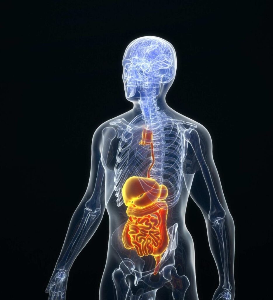
Gastroenterology
Gastroenterology is the branch of medicine focused on the digestive system and its disorders. Diseases affecting the gastrointestinal tract, which include the organs from mouth into anus, along the alimentary canal, are the focus of this speciality.
Sub specialities
- Proctology
- Hepatology
- Interventional Endoscopy
Common Condition / Illness
Select Disease
Hernia
- Hernia
- Obesity
- Chronic Pancreatitis
- Gallstones
- Haemorrhoids
- Anal Fistulas
- Anal fissure
- Pilonidal Sinus
- Appendicitis
Overview
A hernia occurs when an organ pushes through an opening in the muscle or tissue that holds it in place.
Types of hernia:
- Inguinal hernia-Inguinal hernias are the most common type of hernia. They occur when the intestines push through a weak spot or tear in the lower abdominal wall, often in the inguinal canal.
- Hiatal hernia-A hiatal hernia occurs when part of your stomach protrudes up through the diaphragm into your chest cavity.
- Umbilical hernia-Umbilical hernias can affect children and babies. They occur when the intestines bulge through the abdominal wall near the belly button.
- Ventral hernia-A ventral hernia happens when tissue bulges through an opening in the muscles of your abdomen.
Symptoms
- Fever
- Chest pain
- Shortness of breath
- Bloody, black or very dark colored stool
- Difficulty swallowing
- Severe or persistent abdominal pain
- Vomiting, especially if your vomit is bloody or looks like coffee grounds
- Noticeable lump or bulge
- Discomfort or pain.
- Heartburn
- Acid Reflux
- Regurgitation of food or liquids
Procedures
- Endoscopy
- Open Hernia Surgery
- Laparoscopic Hernia Surgery
Endoscopy is a nonsurgical procedure performed in order to look into a person’s digestive tract. Using an endoscope, a flexible tube with a light and camera attached to it, we can view pictures of the digestive tract on a TV monitor.
Colonoscopy—colonoscopy is considered to be the best way to screen the colon for colon cancer and related symptoms. It covers the rectum, the entire length of the large intestine, as well as the lower part of the small intestine (called the small bowel).
Endoscopic Ultrasound (EUS)—endoscopic ultrasound is used to examine the upper or lower GI tract. It is a great way to examine growths previously detected in the body, as the ultrasound technology will show how deep into the tissue the growth has spread and help doctors figure out the best form of treatment accordingly, whether it is biopsy or chemotherapy. EUS is often used to examine a patient’s gallbladder, bile duct and pancreas.
Sigmoidoscopy—flexible sigmoidoscopy is similar to colonoscopy, but is limited to the sigmoid colon and the rectal area of the body only.
Upper Endoscopy—also called an EGD or an esophagogastroduodenoscopy, an upper endoscopy covers the esophagus, stomach and duodenum (uppermost section of the small intestine). It is often performed to diagnose underlying conditions causing abdominal pain, vomiting and dysphagia (difficulty swallowing).
Endoscopic Retrograde Cholangiopancreatography (ERCP)—ERCP is an endoscopic procedure of the liver and pancreas.
Risks
Risks of endoscopy may include:
- Over-sedation, although sedation is not always necessary
- Feeling bloated for a short time after the procedure
- Mild cramping
- A numb throat for a few hours due to the use of local anaesthetic
- Infection of the area of investigation
- Persistent pain in the area of the endoscopy
- Perforation or tear of the lining of the stomach or esophagus occurs in 1 in every 2,500-11,000 cases
- Internal bleeding, usually minor and sometimes treatable by endoscopic cauterization
- Complications related to pre-existing conditions
The surgeon makes an incision (cut) near the hernia and returns the bulging tissue back into the body.
Risks
- Sensory loss
- Hyperesthesia
- Chronic inguinal pain
- Mesh-related problems, Hydrocele
- Testicular pain & testicular swelling
- Atrophy
- Recurrence of hernia.
Multiple, tiny incisions are made around the hernia site to allow for the insertion of long, thin surgical tools. One tool has a camera attached to it, so the surgeon can view images that are projected onto a TV screen. Tools are then used to repair the hernia in the same way as with open surgery.
Risks
- Incisional complications
- Infections
- Bleeding
- Bladder and urinary complications
- Acute and chronic pain complications
- Spermatic cord complications
- Seromas recurrences
- GI complications
- Complications related to general anesthesia.
Overview
Obesity is a complex disease involving an excessive amount of body fat. Obesity isn’t just a cosmetic concern. It’s a medical problem that increases the risk of other diseases and health problems, such as heart disease, diabetes, high blood pressure and certain cancers.
BMI Weight status
Below 18.5 Underweight
18.5-24.9 Normal
25.0-29.9 Overweight
30.0 and higher Obesity
Symptoms
- Your body mass index (BMI) is 40 or higher (extreme obesity).
- Your BMI is 35 to 39.9 (obesity), and you have a serious weight-related health problem, such as type 2 diabetes, high blood pressure or severe sleep apnea.
Procedure
Gastric Bypass Surgery
Gastric bypass and other weight-loss surgeries — known collectively as bariatric surgery — involve making changes to your digestive system to help you lose weight. Bariatric surgery is done when diet and exercise haven’t worked or when you have serious health problems because of your weight.
Risks
- Excessive bleeding
- Infection
- Adverse reactions to anesthesia
- Blood clots
- Lung or breathing problems
- Leaks in your gastrointestinal system
- Death (rare)
Longer term risk
- Bowel obstruction
- Dumping syndrome, causing diarrhea, nausea or vomiting
- Gallstones
- Hernias
- Low blood sugar (hypoglycemia)
- Malnutrition
- Stomach perforation
- Ulcers
- Vomiting
- Death (rare)
Overview
Chronic pancreatitis is a long-standing inflammation of the pancreas that alters the organ’s normal structure and functions. It can present as episodes of acute inflammation in a previously injured pancreas, or as chronic damage with persistent pain or malabsorption
Symptoms
- Upper abdominal pain
- Abdominal pain that radiates to your back
- Tenderness when touching the abdomen
- Fever
- Rapid pulse
- Nausea
- Vomiting
Procedures
- Peustow’s Operation
- Whipple Procedure (Pacreaticduodenectomy/Pancreatoduodenectomy)
The pancreatic duct is opened up from head to tail. Then the small intestine is attached directly to the pancreatic duct using sutures. This results in the pancreatic juice directly draining into the small intestine
Risk
- Excessive bleeding
- Infection
- Ulcers
- Vomiting
The Whipple procedure involves removing the head of the pancreas, the duodenum, the proximal jejunum, gallbladder and distal stomach.
Risks
- Bleeding at the surgical areas
- Infection of the incision area or inside your abdomen
- Delayed emptying of the stomach, which may make it difficult to eat or to keep food down temporarily
- Leakage from the pancreas or bile duct connection
- Diabetes, temporary or permanent
Overview
Gallstones are hardened deposits of digestive fluid that can form in your gallbladder. Your gallbladder is a small, pear-shaped organ on the right side of your abdomen, just beneath your liver. The gallbladder holds a digestive fluid called bile that’s released into your small intestine.
Gallstones range in size from as small as a grain of sand to as large as a golf ball.
Symptoms
- Sudden and rapidly intensifying pain in the upper right portion of your abdomen.
- Sudden and rapidly intensifying pain in the center of your abdomen, just below your breastbone.
- Back pain between your shoulder blades.
- Pain in your right shoulder.
- Nausea or vomiting.
Procedure
Cholecystectomy ( Gallbladder Removal)
Cholecystectomy is a surgical procedure to remove your gallbladder.
- A pear-shaped organ that sits just below your liver on the upper right side of your abdomen. Your gallbladder collects and stores bile
- A digestive fluid produced in your liver.
Cholecystectomy may be necessary if you experience pain from gallstones that block the flow of bile. Cholecystectomy is a common surgery, and it carries only a small risk of complications. In most cases, you can go home the same day of your cholecystectomy.
Cholecystectomy is most commonly performed by inserting a tiny video camera and special surgical tools through four small incisions to see inside your abdomen and remove the gallbladder. Doctors call this laparoscopic cholecystectomy. In some cases, one large incision may be used to remove the gallbladder. This is called an open cholecystectomy.
Overview
Haemorrhoids (HEM-uh-roids), also called piles, are swollen veins in your anus and lower rectum, similar to varicose veins. Haemorrhoids can develop inside the rectum (internal haemorrhoids) or under the skin around the anus (external haemorrhoids).
Nearly three out of four adults will have haemorrhoids from time to time. Haemorrhoids have a number of causes, but often the cause is unknown.
Symptoms
External Haemorrhoids
- Itching or irritation in your anal region
- Pain or discomfort
- Swelling around your anus
- Bleeding
Internal Haemorrhoids
- Painless bleeding during bowel movements. You might notice small amounts of bright red blood on your toilet tissue or in the toilet.
- A haemorrhoid to push through the anal opening (prolapsed or protruding haemorrhoid), resulting in pain and irritation.
Procedures
- Excisional Haemorrhoidectomy
- Stapled hemorrhoidopexy
This is a surgery to remove haemorrhoids. It is done under conventional anaesthetic for the most part although general may also be used. The procedure involves making incisions around the anus. This allows the surgeon to cut out the haemorrhoids. It gives patients relief from pain, swelling, bleeding and itching caused by the haemorrhoids. However, the incisions are in the sensitive place and require stitches, causing pain in itself. It is usually done as an outpatient procedure and patients go home the same day.
Risk
- Pain
- Bleeding
- Infection
- Urine retention
- Abscess (a localized collection of pus)
- Fistula formation (an abnormal communication path between the anus and rectum),
- Loss of bowel control and
- Anal stenosis (abnormal narrowing of the anus)
Stapled hemorrhoidopexy, is a surgical procedure that involves the cutting and removal of Anal Hemorhoidal Vascular Cushion whose function is to help to seal stools and create continence. Procedure also removes abnormally enlarged hemorrhoidal tissue, followed by the repositioning of the remaining hemorrhoidal tissue back to its normal anatomic position.
Risk
- Pain
- Bleeding
- Infection
- Urine retention
- Abscess (a localised collection of pus)
- Fistula formation (an abnormal communication path between the anus and rectum)
- Loss of bowel control
- Anal stenosis (abnormal narrowing of the anus)
Overview
Anal fistula is the medical term for an infected tunnel that develops between the skin and the muscular opening at the end of the digestive tract (anus).
Most anal fistulas are the result of an infection that starts in an anal gland. This infection results in an abscess that drains spontaneously or is drained surgically through the skin next to the anus. The fistula then forms a tunnel under the skin and connects with the infected gland.
Based on their location around the sphincter, anal fistulas are classified into:
- Superficial fistula
- Intersphincteric fistula
- Transsphincteric fistula
- Supraspincteric fistula
- Extrasphincteric fistula
Symptoms
- Anorectal pain, swelling, redness & tenderness.
- Fever.
- Pressure while defecating, cough, sitting.
- Constipation or pain associated with bowel movements.
- Painful urination.
- Foul-smelling discharge from perianal skin.
- Sometimes, rectal bleeding.
Procedures
- Fistuotomy
- Seton Surgery
Fistula Surgery, also known by the name of fistuotomy can be described as a medical procedure specially designed to treat fistula present in the anal.
The primary goal of this treatment is to remove build-up fluids like puss out of small openings developed between anal skin and the end of the anus. The surgical procedure will aid the patient to close up and heal the wounds of the anus fast caused by previous injury, infection, or inflammation.
Risk
- Losing control of one’s bowel.
- Taking a long time for the wound to heal.
- The fistula coming back.
- Narrowing of the anal canal, making it difficult to have a bowel movement.
Setons are used to treat anal fistulas. A fistula is an abnormal tunnel that connects two structures. An anal fistula is an abnormal tunnel that forms between the anus and the surrounding skin. The fistula internal opening is in the anus and the external opening is typically on the skin around the anus or on the buttocks.
If the external opening of the fistula closes, the bacteria inside the fistula tunnel may multiply and form an abscess. An abscess is an infected pocket of fluid, which causes significant pain. Surgeons place a seton (a thin rubber drain that goes through the tunnel) to keep the fistula tract open, which then prevents abscess formation. Usually a second surgical procedure is required to close the fistula, after the seton procedure.
Risk
- Skin irritation around the anus.
- Throbbing pain.
- Unpleasant smelling discharge near the anus.
- Passing blood with a bowel movement.
- Swelling and redness around the anus.
- A high temperature, if an abscess is also present.
- Difficulty controlling bowel movements.
Overview
An anal fissure is a small tear in the thin, moist tissue (mucosa) that lines the anus. An anal fissure may occur when you pass hard or large stools during a bowel movement. Anal fissures typically cause pain and bleeding with bowel movements. You also may experience spasms in the ring of muscle at the end of your anus (anal sphincter).
Anal fissures are very common in young infants but can affect people of any age.
Symptoms
- Pain, sometimes severe, during bowel movements.
- Pain after bowel movements that can last up to several hours.
- Bright red blood on the stool or toilet paper after a bowel movement.
- A visible crack in the skin around the anus.
- A small lump or skin tag on the skin near the anal fissure.
Procedure
Sphincterotomy
This is a surgical procedure, where the doctor makes a small cut in the anal sphincter. Thereby, relaxing the muscle and letting the fissure cure.
Risk
- Temporary incontinence.
- Hemorrhage.
- Perianal abscess.
Overview
Pilonidal sinus is a small hole or channel under the skin and has pus or inflamed fluid accumulation, which may also contain blood. This happens in the cleft, at the lower back or the top of the buttocks. Pilonidal cyst or the sinus may also have hair or dirt accumulation, which may cause severe pain and foul-smelling pus or bloody discharge.
People with regular sitting jobs are at a high risk of developing pilonidal sinus or cyst. It is more common in men, as compared to women. It happens when the hair at the top of the cleft (of the buttocks) gets pushed inside the body, causing dirt to push inside. The condition gets very painful at this stage. In a lot of cases, pilonidal sinus develops from an abscess.
Symptoms
- Pus or blood draining from the abscess, causing a foul odor.
- Blood from the opening of the skin.
- Pain when sitting or standing.
- Pain while sitting.
- Hair Protruding from the lesion.
Procedure
Pilonidal Sinus Surgery
The purpose of surgery is to remove the infection and to prevent another problem pilonidal sinus. If the sinus is very small, it can be completely removed. The wound is closed with stitches. If the sinus is too big to stitch or if there is an infection the wound is not stitched closed. The wound is left open. This allows the skin to fill in from the inside out.
Risk
- Cyst recurrence
- Skin cancer
- Pilonidal abscess starts leaking the foul-smelling liquid and cause the patient pain in the area.
- Chronic pilonidal cystinfections
Overview
Appendicitis is an inflammation of the appendix, a finger-shaped pouch that projects from your colon on the lower right side of your abdomen.
Appendicitis causes pain in your lower right abdomen. However, in most people, pain begins around the navel and then moves. As inflammation worsens, appendicitis pain typically increases and eventually becomes severe.
Although anyone can develop appendicitis, most often it occurs in people between the ages of 10 and 30.
Symptoms
- Sudden pain that begins on the right side of the lower abdomen
- Sudden pain that begins around your navel and often shifts to your lower right abdomen
- Pain that worsens if you cough, walk or make other jarring movements
- Nausea and vomiting
- Loss of appetite
- Low-grade fever that may worsen as the illness progresses
- Constipation or diarrhea
- Abdominal bloating
- Flatulence
Procedure
Appendectomy
Appendectomy can be performed as open surgery using one abdominal incision about 2 to 4 inches (5 to 10 centimeters) long (laparotomy). Or the surgery can be done through a few small abdominal incisions (laparoscopic surgery). During a laparoscopic appendectomy, the surgeon inserts special surgical tools and a video camera into your abdomen to remove your appendix.
Risk
- Bleeding
- Infection
- Injury to nearby organs
- Blocked bowels
Urology & Andrology
Andrology is a branch of medicine that deals with medical and surgical treatments of the male reproductive organs.
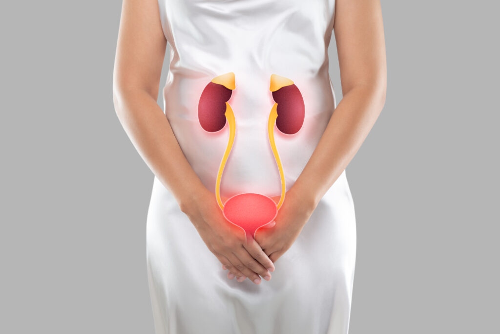
Urology is the branch of medicine which deals with the ailments of the kidneys, bladder and the urinary tract
Sub specialities
- Paediatric Urology
- Uro-oncology
- Uro-gynaecology
- Neuro-urology (Nervous System control of Genitourinary Organs)
Common Condition / Illness
Select Disease
Kidney Stones
- Kidney Stones
- Erectile Dysfunction (ED)
- Enlarged Prostate (BPH)
- Hydrocele
- Ureteral Stricture
- Bladder Stones
- Male Infertility
- Undescended Testicle
Overview
A kidney stone is a hard object that is made from chemicals in the urine. There are four types of kidney stones: calcium oxalate, uric acid, struvite, and cystine. A kidney stone may be treated with shockwave lithotripsy, uteroscopy, percutaneous nephrolithomy or nephrolithotripsy.
Symptoms
- Severe pain on either side of your lower back
- More vague pain or stomach ache that doesn’t go away
- Blood in the urine
- Nausea or vomiting
- Fever and chills
- Urine that smells bad or looks cloudy
Procedures
- Retrograde Intrarenal Surgery (RIRS) for Kidney Stone
- Percutaneous Nephrolithotomy
RIRS is performed to remove stones without making any incisions on the kidney while using a laser and a viewing tube called a fiberoptic endoscope that goes through the urethra into the kidney. It is performed under general, local or spinal anesthesia. This procedure requires a specialized urologist who is specifically trained in RIRS.
To perform this procedure, the scope is placed through the urethra, into the ureter and finally into the urine-collecting part of the kidney. The scope is therefore moved retrograde, i.e. up the urinary tract system to within the kidney, i.e. intrarenal. Once the scope is in place, the doctor can see the stone and can proceed to manipulate or crush it by an ultrasound probe or be evaporated by a laser probe or even grabbed by small forceps.
Risk
- Fever
- Flank pain
- Urinary infection
- Transient hematuria
- Acute urinary retention
- Fornix rupture
- Ureter avulsion
- Bleeding
- Sepsis
Percutaneous nephrolithotomy is typically recommended in the following situations
Large kidney stones are blocking more than one branch of the collecting system of the kidney (known as staghorn kidney stones)
Kidney stones are larger than 0.8 inch (2 centimeters) in diameter. Large stones are in the ureter
- Other therapies have failed
Risk
- Bleeding
- Infection
- Injuries to the kidney or other organs
- Incomplete stone removal
- Other therapies have failed.
- Large stones are in the ureter
- Kidney stones are larger than 0.8 inch (2 centimeters) in diameter
Overview
Erectile Dysfunction (impotence) is the inability to get and keep an erection firm enough for sex.
Having erection trouble from time to time isn’t necessarily a cause for concern. If erectile dysfunction is an ongoing issue, however, it can cause stress, affect your self-confidence and contribute to relationship problems.
Symptoms
- Trouble getting an erection
- Trouble keeping an erection
- Reduced sexual desire
Procedure
Penile Prosthesis
Penile implants are devices placed inside the penis to allow men with erectile dysfunction (ED) to get an erection. Penile implants are typically recommended after other treatments for ED fail.
There are two main types of penile implants, semirigid and inflatable.
Risk
- Uncontrolled bleeding after the surgery; this condition may require an additional surgery
- Infection
- Scar tissue formation
- Erosion (of implant)
- Pump or reservoir displacement
- Mechanical failure
Overview
Benign prostatic hyperplasia (BPH) also called prostate gland enlargement — is a common condition as men get older. An enlarged prostate gland can cause uncomfortable urinary symptoms, such as blocking the flow of urine out of the bladder. It can also cause bladder, urinary tract or kidney problems.
Symptoms
- Frequent or urgent need to urinate
- Increased frequency of urination at night (nocturia)
- Difficulty starting urination
- Weak urine stream or a stream that stops and starts
- Dribbling at the end of urination
- Inability to completely empty the bladder
Procedures
- Transurethral Microwave Thermotherapy (TUMT)
- Transurethral Needle Ablation (TUNA)
Transurethral Microwave Thermotherapy (TUMT) is an outpatient procedure to treat urinary symptoms caused by an enlarged prostate, a condition known as benign prostatic hyperplasia (BPH). A small microwave antenna is inserted through the tip of the penis into the tube that carries urine from the bladder (urethra). The doctor extends the antenna until it reaches the area of the urethra surrounded by the prostate. The antenna emits a dose of microwave energy that heats and destroys the excess prostate tissue that is blocking urine flow.
Risk
- New onset or worsening urinary symptoms
- Temporary difficulty urinating
- Urinary tract infection
Transurethral needle ablation, also known as TUNA or radiofrequency ablation, is a minimally invasive treatment option used to treat benign prostatic hyperplasia.
During the procedure, radiofrequency needles are placed through the urethra into the area of the prostate that is pressing on the urethra. Radio waves are sent through the needles, into the prostate tissue to destroy prostate tissue. As a result, the prostate shrinks, which allows urine to flow out the urethra.
Risk
- Chronic inflammation of the prostate, which could cause painful urination or frequent need to urinate
- Difficulty urinating for a few days after the procedure
- Urinary tract infection
- Erectile dysfunction, while rare, can occur
- Need to be treated again
Overview
A hydrocele (HI-droe-seel) is a type of swelling in the scrotum that occurs when fluid collects in the thin sheath surrounding a testicle. Hydrocele is common in newborns and usually disappears without treatment by age 1. Older boys and adult men can develop a hydrocele due to inflammation or injury within the scrotum.
A hydrocele usually isn’t painful or harmful and might not need any treatment. But if you have scrotal swelling, see your doctor to rule out other causes.
Symptoms
Usually, the only indication of a hydrocele is a painless swelling of one or both testicles.
Adult men with a hydrocele might experience discomfort from the heaviness of a swollen scrotum. Pain generally increases with the size of the inflammation.
Procedure
Hydrocelectomy
Hydrocelectomy is surgery that is done to remove or repair a hydrocele. A hydrocele is a fluid-filled sac surrounding a testicle that causes swelling in the scrotum (the pouch that holds the testicles).
Risk
- Redness or a feeling of warmth at the surgical site
- Increasing pain
- Bad-smelling fluid seeping from the surgical wound
- Increasing swelling
- Fever
Overview
A urethral (u-REE-thrul) stricture involves scarring that narrows the tube that carries urine out of your body (urethra). A stricture restricts the flow of urine from the bladder and can cause a variety of medical problems in the urinary tract, including inflammation or infection.
Symptoms
- Decreased urine stream
- Incomplete bladder emptying
- Spraying of the urine stream
- Difficulty, straining or pain when urinating
- Increased urge to urinate or more-frequent urination
- Urinary tract infection
Procedures
- Ureteroplasty
- Nephrostomy
Urethroplasty is a surgical procedure where the urethra is reconstructed to treat various stricture problems.The surgery may be done through several small cuts (incisions). This type of surgery is called laparoscopy. Or it will be done through one larger cut. This is called open surgery. Laparoscopy can’t be used in all cases. In some cases, your surgeon may start the surgery using laparoscopy but must change to open surgery for safety reasons.
Risk
- Bleeding (may require a blood transfusion)
- Infection
- Urine leakage from the ureter or bladder
- Stricture returning after surgery
- Kidney damage
- Blood clots
- Risks of anesthesia (the anesthesiologist or nurse anesthetist will discuss these with you)
A nephrostomy is a procedure to drain urine from your kidney using a catheter (tube). Urine normally drains from your kidneys into your bladder through small muscular tubes (ureters).
Risk
- Bleeding
- Infection
- Allergic reaction
- Making a hole in nearby structures with the needle
- Leaking urine
- Failed nephrostomy
- Blocked nephrostomy
- Radiation exposure
Overview
Bladder stones are hard masses of minerals in your bladder. They develop when the minerals in concentrated urine crystallize and form stones. This often happens when you have trouble completely emptying your bladder.
Small bladder stones may pass without treatment, but sometimes bladder stones need medications or surgery.
Symptoms
- Lower abdominal pain
- Pain during urination
- Frequent urination
- Difficulty urinating or interrupted urine flow
- Blood in the urine
- Cloudy or unusually dark-colored urine.
Procedure
Cystoscopy
Cystoscopy is a procedure that allows your doctor to examine the lining of your bladder and the tube that carries urine out of your body (urethra). A hollow tube (cystoscope) equipped with a lens is inserted into your urethra and slowly advanced into your bladder.
Risk
- Infection. Rarely, cystoscopy can introduce germs into your urinary tract, causing an infection. Risk factors for developing a urinary tract infection after cystoscopy include advanced age, smoking and unusual anatomy in your urinary tract.
- Bleeding. Cystoscopy might cause some blood in your urine. Serious bleeding occurs rarely.
- Pain. After the procedure, you might experience abdominal pain and a burning sensation when you urinate. These symptoms are generally mild and gradually get better after the procedure.
Overview
Nearly 1 in 7 couples is infertile, which means they haven’t been able to conceive a child even though they’ve had frequent, unprotected sexual intercourse for a year or longer. In up to half of these couples, male infertility plays at least a partial role.
Male infertility can be caused by low sperm production, abnormal sperm function or blockages that prevent the delivery of sperm. Illnesses, injuries, chronic health problems, lifestyle choices and other factors may contribute to male infertility.
Symptoms
- Problems with sexual function — for example, difficulty with ejaculation or small volumes of fluid ejaculated, reduced sexual desire, or difficulty maintaining an erection (erectile dysfunction)
- Pain, swelling or a lump in the testicle area
- Recurrent respiratory infections
- Inability to smell
- Abnormal breast growth (gynecomastia)
- Decreased facial or body hair or other signs of a chromosomal or hormonal abnormality
- A lower than normal sperm count (fewer than 15 million sperm per milliliter of semen or a total sperm count of less than 39 million per ejaculate)
Procedure
Vasectomy
Vasectomy is a form of male birth control that cuts the supply of sperm to your semen. It’s done by cutting and sealing the tubes that carry sperm. Vasectomy is a safe and effective birth control choice for men who are certain they don’t want to father a child in the future.
Risk
- Chronic pain, which can happen for 1% to 2% of people who have surgery.
- Fluid buildup in the testicle, which can cause a dull ache that gets worse with ejaculation.
- Inflammation caused by leaking sperm (granuloma).
- Pregnancy, in the event that your vasectomy fails, which is rare.
- An abnormal cyst (spermatocele) that develops in the small, coiled tube located on the upper testicle that collects and transports sperm (epididymis).
- A fluid-filled sac (hydrocele) surrounding a testicle that causes swelling in the scrotum.
Overview
An undescended testicle (cryptorchidism) is a testicle that hasn’t moved into its proper position in the bag of skin hanging below the penis (scrotum) before birth. Usually just one testicle is affected, but about 10 percent of the time both testicles are undescended.
An undescended testicle is uncommon in general, but common among baby boys born prematurely.
Symptoms
Not seeing or feeling a testicle where you would expect it to be in the scrotum is the main sign of an undescended testicle.
Orchidopexy
Orchidopexy, or orchiopexy, is a surgical procedure which is performed in order to fix an undescended testicle into the scrotum. Undescended testicles generally move to the scrotum by the time your baby is 3 to 6 months old. If the testicles do not descend by this point, they will generally need to be surgically corrected. If orchidopexy is not performed, the chances of fertility problems or testicular cancer increases later in life.
Risk
- Blood clots in the scrotum.
- Damage to the structures and/or tissues of the testicle.
- Inadequate blood supply, which can lead to atrophy (shrinkage).
Nephrology
Nephrology is the subspecialty of internal medicine that focuses on the diagnosis and treatment of diseases of the kidney. Kidneys are essential for filtering out waste products and excess water from the body. They are also vital for retaining the fluid intake, electrolytes that may be altered by numerous conditions or medicines.
Sub specialities
- Interventional Nephrology
- Onco-nephrology
- Paediatric Nephrology
- Kidney Transplantation

Common Condition / Illness
Select Disease
Chronic kidney disease
- Chronic Kidney Disease
- Amyloidosis
- Glomerulonephritis
Overview
Chronic Kidney Disease, also called chronic kidney failure, involves a gradual loss of kidney function. Your kidneys filter wastes and excess fluids from your blood, which are then removed in your urine. Advanced chronic kidney disease can cause dangerous levels of fluid, electrolytes and wastes to build up in your body.
Symptoms
- Nausea
- Vomiting
- Loss of appetite
- Fatigue and weakness
- Sleep problems
- Urinating more or less
- Decreased mental sharpness
- Muscle cramps
- Swelling of feet and ankles
- Dry and itchy skin
- High blood pressure (hypertension) that’s difficult to control
- Shortness of breath, if fluid builds up in the lungs
- Chest pain, if fluid builds up around the lining of the heart
Procedure
- Dialysis
- Kidney Transplant
The kidneys are responsible for purifying the blood by removing excess fluid and waste from it. When they do not work properly or fail, it is a life-threatening condition. In these cases a machine is used to do the work of the kidneys. It purifies the blood through filtration and makes it viable for the body. The process is called dialysis. There are two kinds of dialysis – haemodialysis and peritoneal dialysis.
Haemodialysis
Haemodialysis is the more common form of dialysis. It makes use of an ‘artificial kidney’ or machine that is known as a haemodialyser to filter chemicals and waste from the blood.
Peritoneal Dialysis
This is a less common form of dialysis. It utilises the lining of the abdominal region as a filter to clean blood. The advantages of it are that it can be carried out while the patient is asleep to at work or even during the course of daily activity. Prior to the first session, surgery is required to create access into the region. A small incision is made (usually to the side of the belly button) and a catheter is inserted into the peritoneal cavity. A cleaning solution, called dialysate, is passed through the catheter into the region. The excess fluid and waste which is in the blood passes through the lining of the peritoneal cavity and is drawn into the dialysate solution. This is allowed to happen to for about four to six hours, after which the solution is drained. Along with it, comes the waste and excess fluid.
Risk: Hemodialysis
- Low blood pressure (hypotension)
- Muscle cramps
- Anemia
- Bone diseases
- High blood pressure (hypertension)
- Fluid overload
- Amyloidosis
- Depression
Risk: Peritoneal Dialysis
- Weight gain
- Inadequate dialysis
- Hernia
A Kidney transplant is a surgical procedure to place a healthy kidney from a living or deceased donor into a person whose kidneys no longer function properly
Types:
A preemptive kidney transplant is when you receive a kidney transplant before your kidney function deteriorates to the point of needing dialysis to replace the normal filtering function of the kidneys.
A deceased-donor kidney transplant is when a kidney from someone who has recently died is removed with consent of the family or from a donor card and placed in a recipient whose kidneys have failed and no longer function properly and is in need of kidney transplantation.
A living-donor kidney transplant is when a kidney from a living donor is removed and placed into a recipient whose kidneys no longer function properly.
Risk
- Blood clots and bleeding
- Leaking from or blockage of the tube (ureter) that links the kidney to the bladder Infection
- Failure or rejection of the donated kidney
- An infection or cancer that can be transmitted with the donated kidney
- Death, heart attack and stroke
Overview
Amyloidosis (am-uh-loi-DO-sis) is a rare disease that occurs when an abnormal protein, called amyloid, builds up in your organs and interferes with their normal function.
Amyloid isn’t normally found in the body, but it can be formed from several different types of protein. Organs that may be affected include the heart, kidneys, liver, spleen, nervous system and digestive tract.
Symptoms
- Swelling of your ankles and legs
- Severe fatigue and weakness
- Shortness of breath with minimal exertion
- Unable to lie flat in bed due to shortness of breath
- Numbness, tingling or pain in your hands or feet, especially pain in your wrist (carpal tunnel syndrome)
- Diarrhea, possibly with blood, or constipation
- Unintentional weight loss of more than 10 pounds (4.5 kilograms)
- An enlarged tongue, which sometimes looks rippled around its edge
- Skin changes, such as thickening or easy bruising, and purplish patches around the eyes
- An irregular heartbeat
- Difficulty swallowing
Procedures
Overview
Glomerulonephritis (gloe-mer-u-low-nuh-FRY-tis) is inflammation of the tiny filters in your kidneys (glomeruli). Glomeruli remove excess fluid, electrolytes and waste from your bloodstream and pass them into your urine. Glomerulonephritis can come on suddenly (acute) or gradually (chronic).
Symptoms
- Pink or cola-colored urine from red blood cells in your urine (hematuria)
- Foamy urine due to excess protein (proteinuria)
- High blood pressure (hypertension)
- Fluid retention (edema) with swelling evident in your face, hands, feet and abdomen
Procedures
- Therapeutic Plasma Exchange (TPE)
Therapeutic Plasma Exchange (TPE) is a procedure in which the patient’s blood is passed through an apheresis machine, where the filtered plasma is removed and discarded with reinfusion of red blood cells along with replacement fluid such as plasma or albumin in to the patient.
Risk
- Disorders of the lymphatic system
- Blood disorders
- Family or personal history of cancer or malignant tumors
- Exposure to chemicals such as toxic hydrocarbon solvents
- Recurring strep infections or skin abscesses
- Viral infections
- Heart infections
- Specific diseases such as amyloidosis, diseases of the blood vessels such as vasculitis
Cardiothoracic Surgery
Cardiothoracic surgery is the specialty involved with the surgical treatment of diseases affecting organs within the thorax (the chest), principally the heart, lungs and oesophagus.
Sub specialities
- Paediatric Cardiac Surgery
- Thoracic Surgery
- Heart Transplant

Common Condition / Illness
Select Disease
Myocardial Infraction (Heart Attack)
- Myocardial Infraction (Heart Attack)
- Coronary Artery Disease
- Heart Valve Disease
- Aortic Aneurysm
- Congenital Heart Defect
- Aneurysms
Overview
A heart attack occurs when the flow of blood to the heart is blocked. The blockage is most often a buildup of fat, cholesterol and other substances, which form a plaque in the arteries that feed the heart (coronary arteries).
Sometimes, a plaque can rupture and form a clot that blocks blood flow. The interrupted blood flow can damage or destroy part of the heart muscle.
Symptoms
- Pressure, tightness, pain, or a squeezing or aching sensation in your chest or arms that may spread to your neck, jaw or back
- Nausea, indigestion, heartburn or abdominal pain
- Shortness of breath
- Cold sweat
- Fatigue
- Lightheadedness or sudden dizziness
Procedures
- Coronary angioplasty and stenting
- Coronary bypass surgery
Coronary angioplasty (AN-jee-o-plas-tee), also called percutaneous coronary intervention, is a procedure used to open clogged heart arteries. Angioplasty uses a tiny balloon catheter that is inserted in a blocked blood vessel to help widen it and improve blood flow to the heart.
Angioplasty is often combined with the placement of a small wire mesh tube called a stent. The stent helps prop the artery open, decreasing its chance of narrowing again. Most stents are coated with medication to help keep the artery open (drug-eluting stents). Rarely, bare-metal stents are used.
Risk
- Re-narrowing of your artery
- Blood clots
- Bleeding
Coronary bypass surgery redirects blood around a section of a blocked or partially blocked artery in your heart. The procedure involves taking a healthy blood vessel from your leg, arm or chest and connecting it below and above the blocked arteries in your heart. With a new pathway, blood flow to the heart muscle improves.
Risk
- Bleeding
- An irregular heart rhythm
- Infections of the chest wound
- Memory loss or trouble thinking clearly, which often improves within six to 12 months
- Kidney problems
- Stroke
- Heart attack, if a blood clot breaks loose soon after surgery
Overview
Coronary artery disease develops when the major blood vessels that supply your heart become damaged or diseased. Cholesterol-containing deposits (plaques) in your coronary arteries and inflammation are usually to blame for coronary artery disease.
The coronary arteries supply blood, oxygen and nutrients to your heart. A buildup of plaque can narrow these arteries, decreasing blood flow to your heart. Eventually, the reduced blood flow may cause chest pain (angina), shortness of breath, or other coronary artery disease signs and symptoms. A complete blockage can cause a heart attack.
Symptoms
- Chest pain (angina)
- Shortness of breath
- Heart attack
Procedures
- Coronary artery bypass graft surgery (CABG)
Coronary artery bypass graft surgery (CABG) is a procedure used to treat coronary artery disease. Coronary artery disease (CAD) is the narrowing of the coronary arteries – the blood vessels that supply oxygen and nutrients to the heart muscle. CAD is caused by a build-up of fatty material within the walls of the arteries. This build-up narrows the inside of the arteries, limiting the supply of oxygen-rich blood to the heart muscle.
Risk
- Bleeding during or after the surgery
- Blood clots that can cause heart attack, stroke, or lung problems
- Infection at the incision site
- Pneumonia
- Breathing problems
- Pancreatitis
- Kidney failure
- Abnormal heart rhythms
- Failure of the graft
- Death
Overview
In heart valve disease, one or more of the valves in your heart doesn’t work properly.
Your heart has four valves that keep blood flowing in the correct direction. In some cases, one or more of the valves don’t open or close properly. This can cause the blood flow through your heart to your body to be disrupted.
Symptoms
- Whooshing sound (heart murmur) when a doctor is listening to the heart with a stethoscope
- Chest pain
- Abdominal swelling (more common with advanced tricuspid regurgitation)
- Fatigue
- Shortness of breath, particularly when active or lying down
- Swelling of your ankles and feet
- Dizziness
- Fainting
- Irregular heartbeat
Procedures
- Heart Valve Repair/Replacement Surgery
Heart valve surgery is a procedure to treat heart valve disease. Heart valve disease involves at least one of the four heart valves not working properly. Heart valves keep blood flowing in the correct direction through the heart.
The four heart valves are the mitral valve, tricuspid valve, pulmonary valve and aortic valve. Each valve has flaps — called leaflets for the mitral and tricuspid valves and cusps for the aortic and pulmonary valves. These flaps should open and close once during each heartbeat. Valves that don’t open or close properly disrupt blood flow through the heart to the body.
In heart valve surgery, a surgeon repairs or replaces the damaged or diseased heart valve or valves.
Types:
Annuloplasty
Valvuloplasty
Risk
- Bleeding
- Heart attack
- Infection
- Valve dysfunction affecting replaced valves
- Irregular heart rhythm (arrhythmia)
- Stroke
- Death
Overview
An aortic aneurysm is a bulge that occurs in the wall of the major blood vessel (aorta) that carries blood from the heart to the body. Aortic aneurysms can occur anywhere in the aorta and may be tube-shaped (fusiform) or round (saccular).
Aortic aneurysms include:
Abdominal aortic aneurysm. An abdominal aortic aneurysm occurs along the part of the aorta that passes through the abdomen.
Thoracic aortic aneurysm. A thoracic aortic aneurysm occurs along the part of the aorta that passes through the chest cavity.
Procedures
- Thoracic Endovascular Aortic Repair (TEVAR)
- Endovascular Aneurysm Repair (EVAR)
Thoracic endovascular aortic repair (TEVAR) is a procedure to treat an aneurysm in the upper part of your aorta. The aorta is your body’s largest artery. An aneurysm is a weak, bulging area in the aorta wall. If it bursts (ruptures), it can be deadly.
TEVAR is a minimally invasive surgery. That means it is done with a small cut (incision). With TEVAR, a device called a stent graft is used to reinforce the aneurysm. A stent graft is a metal tube covered in fabric. It helps prevent the aneurysm from bursting.
Risk
- Infection
- Bleeding
- Injury to nearby organs
- Blood clots
- Risks from anesthesia
- Kidney damage from dye used during the X-ray when the stent graft is put in place
- Device or delivery failure
- Blood vessel injury
- Leaking graft
- Paralysis
- The graft moves out of place
- Loss of a leg
- Traditional open surgery may be needed
- Your aortic aneurysm may
- keep growing after surgery
Endovascular aneurysm repair (EVAR). EVAR requires only small incisions in the groin. Using X-ray guidance and specially-designed instruments, the surgeon can repair the aneurysm by inserting a metal mesh coil, called a stent-graft, inside the aorta.
Risk
- Heart attack
- Stroke
- Kidney failure
- Chest problem
- Loss of circulation in the legs or bowel
- Infection in the graft used to replace your aorta
Overview
Congenital heart disease is one or more problems with the heart’s structure that exist since birth. Congenital means that you’re born with the defect. Congenital heart disease, also called congenital heart defect, can change the way blood flows through your heart. Some congenital heart defects might not cause any problems. Complex defects, however, can cause life-threatening complications.
Symptoms
- Abnormal heart rhythms (arrhythmias)
- A bluish tint to the skin, lips and fingernails (cyanosis)
- Shortness of breath
- Tiring quickly upon exertion
- Swelling of body tissue or organs (edema)
Procedures
- Heart Transplant
A heart transplant is an operation in which a diseased, failing heart is replaced with a healthier donor heart. Heart transplant is a treatment that’s usually reserved for people whose condition hasn’t improved enough with medications or other surgeries
For some people who cannot have a heart transplant, another option may be a ventricular assist device (VAD). A ventricular assist device is a mechanical pump implanted in your chest that helps pump blood from the lower chambers of your heart (ventricles) to the rest of your body.
VADs are commonly used as temporary treatments for people waiting for heart transplants.
Risk
- Rejection of the donor heart
- Primary graft failure
- Problems with your arteries
- Medication side effects
- Cancer
- Infection
Overview
An aneurysm is an abnormal bulge or ballooning in the wall of a blood vessel. An aneurysm can burst (rupture), causing internal bleeding and often leading to death. Aneurysms usually don’t cause symptoms, so you might not know you have an aneurysm even if it’s large.
Aneurysms can develop in several parts of your body, including:
The aorta — the major blood vessel carrying blood from your heart to vital organs (aortic aneurysm)
The section of aorta that passes through your abdomen (abdominal aortic aneurysm)
The section of aorta that passes through your chest (thoracic aortic aneurysm)
Blood vessels supplying blood to your brain (brain aneurysm)
Blood vessels in other parts of your body, such as your legs, groin or neck (peripheral aneurysm)
Procedures
- Surgical Ventricular Restoration (SVR)
With this technique the left ventricle is opened through the anteroapical scar and a mannequin device is used to size for appropriate right sizing of the left ventricle. A Dacron patch is then sutured the edges of the scar and the left ventricle closed over the Dacron patch. This technique prevents obliteration of the left ventricular cavity, which was a problem with some of the previous ventricular aneurysmectomy techniques.
Cardiology
Cardiology is a branch of medicine that concerns diseases and disorders of the heart, which may range from congenital defects through to acquired heart diseases
Sub specialities
- Interventional Cardiology
- Cardiac Electrophysiology Study
- Paediatric Cardiology
- Non–invasive Cardiology
- Cardiac Rehabilitation

Common Condition / Illness
Select Disease
Angina
- Angina
- Arteriosclerosis / Atherosclerosis
- Arterial Embolism
- Atrial Septal Defect (ASD)
- Arrhythmias
- Bradycardia
- Cardiomyopathy
- Atrial Fibrillation
Overview
Angina is a type of chest pain caused by reduced blood flow to the heart. Angina (an-JIE-nuh or AN-juh-nuh) is a symptom of coronary artery disease.
Angina, also called angina pectoris, is often described as squeezing, pressure, heaviness, tightness or pain in your chest.
Symptoms
- Dizziness
- Fatigue
- Nausea
- Shortness of breath
- Sweating
Procedures
- Enhanced External Counter Pulsation (EECP)
Enhanced External Counter Pulsation (EECP) is performed as a non-invasive treatment to lower the number and intensity of angina episodes. Treatment is administered through three pairs of external inflatable cuffs that are applied around the lower legs, upper legs and buttocks.
Risk
Occasionally, some patients complain of mild skin abrasion or bruising under the cuffs used to perform the therapy.
Overview
Arteriosclerosis occurs when the blood vessels that carry oxygen and nutrients from your heart to the rest of your body (arteries) become thick and stiff — sometimes restricting blood flow to your organs and tissues. Healthy arteries are flexible and elastic, but over time, the walls in your arteries can harden, a condition commonly called hardening of the arteries.
Atherosclerosis is a specific type of arteriosclerosis.
Symptoms
- If you have atherosclerosis in your heart arteries, you may have symptoms, such as chest pain or pressure (angina).
- If you have atherosclerosis in the arteries leading to your brain, you may have signs and symptoms such as sudden numbness or weakness in your arms or legs, difficulty speaking or slurred speech, temporary loss of vision in one eye, or drooping muscles in your face.
- These signal a Transient Ischemic Attack (TIA), which, if left untreated, may progress to a stroke.
- If you have atherosclerosis in the arteries in your arms and legs, you may have signs or symptoms of peripheral artery disease, such as leg pain when walking (claudication) or decreased blood pressure in an affected limb.
- If you have atherosclerosis in the arteries leading to your kidneys, you develop high blood pressure or kidney failure.
Procedures
- Carotid Endarterectomy
Carotid Endarterectomy is a procedure to treat carotid artery disease. This disease occurs when fatty, waxy deposits build up in one of the carotid arteries. The carotid arteries are blood vessels located on each side of your neck (carotid arteries)
Risk
- Restenosis, or the redevelopment of plaque
- Nerve Damage
- Heart Attack
- Strokes and Mini Strokes
Overview
Arterial embolism refers to a clot (embolus) that has come from another part of the body and causes a sudden interruption of blood flow to an organ or body part.
Symptoms
- Cold arm or leg
- Decreased or no pulse in an arm or leg
- Lack of movement in the arm or leg
- Pain in the affected area
- Numbness and tingling in the arm or leg
- Pale color of the arm or leg (pallor)
- Weakness of an arm or leg
Procedures
- Embolectomy
An embolectomy is surgery to remove an embolus from an artery or vein. An embolus is part of a blood clot that broke free. It can travel through your bloodstream and become stuck in another area.
Risk
- Damage to blood vessels
- Development of another blood clot in the treated blood vessel
- Heart arrhythmias
- Heart attack
- Low blood pressure
- Movement of the blood clot to another area of the body while trying to remove it
- Pulmonary embolism
- Stroke
Overview
An Atrial Septal Defect (ASD) is a hole in the wall (septum) between the two upper chambers of your heart (atria). The condition is present at birth (congenital).
Symptoms
- Shortness of breath, especially when exercising
- Fatigue
- Swelling of legs, feet or abdomen
- Heart palpitations or skipped beats
- Stroke
- Heart murmur, a whooshing sound that can be heard through a stethoscope
Procedures
- Cardiac Catheterization
Cardiac catheterization is a procedure in which a thin, flexible tube (catheter) is guided through a blood vessel to the heart to diagnose or treat certain heart conditions, such as clogged arteries or irregular heartbeats. Cardiac catheterization gives doctors important information about the heart muscle, heart valves and blood vessels in the heart.
Risk
- Bleeding
- Blood clots
- Bruising
- Damage to the artery, heart or the area where the catheter was inserted
- Heart attack
- Infection
- Irregular heart rhythms (arrhythmias)
- Kidney damage
- Stroke
- Allergic reactions to the contrast dye or medication
Overview
A heart arrhythmia (uh-RITH-me-uh) is an irregular heartbeat. Heart rhythm problems (heart arrhythmias) occur when the electrical signals that coordinate the heart’s beats don’t work properly. The faulty signaling causes the heart to beat too fast (tachycardia), too slow (bradycardia) or irregularly.
Heart arrhythmias may feel like a fluttering or racing heart and may be harmless.
In general, heart arrhythmias are grouped by the speed of the heart rate. For example:
Tachycardia (tak-ih-KAHR-dee-uh) is a fast heart. The resting heart rate is greater than 100 beats a minute.
Bradycardia (brad-e-KAHR-dee-uh) is a slow heartbeat. The resting heart rate is less than 60 beats a minute.
Symptoms
- A fluttering in the chest
- A racing heartbeat (tachycardia)
- A slow heartbeat (bradycardia)
- Chest pain
- Shortness of breath
Procedures
- Open Heart Surgery
- Catheter Ablation
During open heart surgery, the chest, including the breast bone, is cut open to expose the heart. This incision is usually eight to 10 inches long, depending on the specific procedure, and gives surgeons direct access to the heart.
There are two ways to perform open-heart surgery:
On-pump
Off-pump
Risk
- Allergic reaction to anesthesia
- Arrhythmias (irregular heartbeat)
- Bleeding
- Damage to surrounding blood vessels or organs like the lungs or kidneys
- Infections
- Stroke
Catheter Ablation is a treatment for cardiac arrhythmias. During ablation, a doctor inserts a catheter (thin, flexible tube) into the heart. A special machine delivers energy through the catheter to tiny areas of the heart muscle that cause the abnormal heart rhythm. This energy “disconnects” the pathway of the abnormal rhythm.
Ablation can also be used to disconnect the electrical pathway between the upper chambers (atria) and lower chambers (ventricles) of the heart.
Risk
- Bruising or swelling at the puncture site.
- Blood clots and strokes
- Phrenic nerve
- Injury
- Esophageal fistula
- Pulmonary vein stenosis
- Cardiac perforation
Overview
Bradycardia (brad-e-KAHR-dee-uh) is a slower than normal heart rate. The hearts of adults at rest usually beat between 60 and 100 times a minute. If you have bradycardia, your heart beats fewer than 60 times a minute.
Bradycardia can be a serious problem if the heart rate is very slow and the heart can’t pump enough oxygen-rich blood to the body. If this happens, you may feel dizzy, very tired or weak, and short of breath. Sometimes bradycardia doesn’t cause symptoms or complications.
Symptoms
- Chest pain
- Confusion or memory problems
- Dizziness or lightheadedness
- Easily tiring during physical activity
- Fatigue
- Fainting (syncope) or near-fainting
- Shortness of breath
Procedures
- Pacemaker Surgery
A pacemaker is a small device that’s placed (implanted) in the chest to help control the heartbeat. It’s used to prevent the heart from beating too slowly. Implanting a pacemaker in the chest requires a surgical procedure.
A pacemaker is also called a cardiac pacing device.
Types
Depending on your condition, you might have one of the following types of pacemakers.
Single chamber pacemaker. This type usually carries electrical impulses to the right ventricle of your heart.
Dual chamber pacemaker. This type carries electrical impulses to the right ventricle and the right atrium of your heart to help control the timing of contractions between the two chambers.
Biventricular pacemaker. Biventricular pacing, also called cardiac resynchronization therapy, is for people who have heart failure and heartbeat problems. This type of pacemaker stimulates both of the lower heart chambers (the right and left ventricles) to make the heart beat more efficiently.
Risk
- Infection near the site in the heart, where the device is implanted
- Swelling, bruising or bleeding at the pacemaker site, especially if you take blood thinners
- Blood clots (thromboembolism) near the pacemaker site
- Damage to blood vessels or nerves near the pacemaker
- Collapsed lung (pneumothorax)
- Blood in the space between the lung and chest wall (haemothorax)
- Movement (shifting) of the device or leads, which could lead to cardiac perforation (rare)
Overview
Cardiomyopathy (kahr-dee-o-my-OP-uh-thee) is a disease of the heart muscle that makes it harder for your heart to pump blood to the rest of your body. Cardiomyopathy can lead to heart failure.
The main types of cardiomyopathy include dilated, hypertrophic and restrictive cardiomyopathy.
Symptoms
- Breathlessness with activity or even at rest
- Swelling of the legs, ankles and feet
- Bloating of the abdomen due to fluid buildup
- Cough while lying down
- Difficulty lying flat to sleep
- Fatigue
- Heartbeats that feel rapid, pounding or fluttering
- Chest discomfort or pressure
- Dizziness, lightheadedness and fainting
Procedures
- Implantable Cardioverter- Defibrillator (ICD) Surgery
An Implantable Cardioverter-Defibrillator (ICD) is a small battery-powered device placed in the chest to detect and stop irregular heartbeats (arrhythmias). An ICD continuously monitors the heartbeat and delivers electric shocks, when needed, to restore a regular heart rhythm. An ICD differs from a pacemaker — an implantable device that can prevent dangerously slow heartbeats.
Risk
- Infection at the implant site
- Swelling, bleeding or bruising
- Blood vessel damage from ICD leads
- Bleeding around the heart, which can be life-threatening
- Blood leaking through the heart valve (regurgitation) where the ICD lead is placed
- Collapsed lung (pneumothorax)
- Movement (shifting) of the device or leads, which could lead to cardiac perforation (rare)
Overview
Atrial Fibrillation (A-fib) is an irregular and often very rapid heart rhythm (arrhythmia) that can lead to blood clots in the heart. A-fib increases the risk of stroke, heart failure and other heart-related complications.
During atrial fibrillation, the heart’s upper chambers (the atria) beat chaotically and irregularly — out of sync with the lower chambers (the ventricles) of the heart.
Symptoms
- Sensations of a fast, fluttering or pounding heartbeat (palpitations)
- Chest pain
- Dizziness
- Fatigue
- Lightheadedness
- Reduced ability to exercise
- Shortness of breath
- Weakness
Procedures
- Maze Procedure
Maze is a surgical procedure used to treat atrial fibrillation. A doctor creates a pattern of scar tissue (the maze) in the upper chambers of the heart by applying heat or cold. Or, the doctor uses a scalpel to make several precise incisions. This method is more complex and takes longer.
Interventional Radiology
Interventional Radiology (IR) is a medical subspecialty that performs various minimally-invasive procedures using medical imaging guidance, such as x-ray fluoroscopy, computed tomography, magnetic resonance imaging, or ultrasound. IR performs both diagnostic and therapeutic procedures through very small incisions or body orifices.
Sub specialities
- Neuro Radiology
- Nuclear Radiology
- Pain Medicine
- Paediatric Radiology

Common Condition / Illness
Select Disease
Arteriovenous malformation (AVM)
- Arteriovenous Malformation (AVM)
- Brain Aneurysms
- Congenital Heart Disease
- Tumor
- Interventions Liver Disorders
Overview
A brain arteriovenous malformation (AVM) is a tangle of abnormal blood vessels connecting arteries and veins in the brain.
The arteries are responsible for taking oxygen-rich blood from the heart to the brain. Veins carry the oxygen-depleted blood back to the lungs and heart. A brain AVM disrupts this vital process.
An arteriovenous malformation can develop anywhere in your body but occurs most often in the brain or spine
Symptoms
- Severe headache
- Weakness, numbness or paralysis
- Vision loss
- Difficulty speaking
- Confusion or inability to understand others
- Severe unsteadiness
Procedures
- Endovascular Embolization
- Stereotactic Radio Surgery (SRS)
Healthcare providers use endovascular coiling, also called endovascular embolization, to block blood flow into an aneurysm. An aneurysm is a weakened area in the wall of an artery. If an aneurysm ruptures, it can cause life-threatening bleeding and brain damage. Preventing blood flow into an aneurysm helps to keep it from rupturing
For endovascular coiling, healthcare providers use a catheter, a long, thin tube inserted into a groin artery. The catheter is advanced into the affected brain artery where the coil is deployed. X-rays help guide the catheter into the artery. The coils are made of soft platinum metal, and are shaped like a spring.
Risk
- Loss of consciousness
- Stroke or transient ischemic attack (TIA, a temporary stroke-like condition)
- Paralysis of one half of the body
- Blood clot
- Bleeding
- An area of swelling caused by a collection of blood (hematoma)
- Loss of the ability or speak or the ability to understand speech (aphasia)
- Infection
- Rupture of unruptured aneurysm
- Higher chance of an aneurysm recurring
Stereotactic radiosurgery (SRS) uses many precisely focused radiation beams to treat tumors and other problems in the brain, neck, lungs, liver, spine and other parts of the body.
It is not surgery in the traditional sense because there’s no incision. Instead, stereotactic radiosurgery uses 3D imaging to target high doses of radiation to the affected area with minimal impact on the surrounding healthy tissue.
Risk
- Fatigue
- Swelling
- Scalp and hair problems
Overview
An aneurysm is an abnormal bulge or ballooning in the wall of a blood vessel. An aneurysm can burst (rupture), causing internal bleeding and often leading to death.
Procedures
- Endovascular embolization (EE) embolization
Endovascular embolization (EE) is an invasive surgical procedure. It’s used to treat abnormal blood vessels found in your brain, as well as other areas of your body.
This procedure is an alternative to open surgery. It blocks blood vessels to cut off blood flow to an affected area.
Risk
- Recurring symptoms
- Bleeding into your brain
- Bleeding at the site of your incision
- Damage to the artery where the catheter is inserted
- Failure of the blocking material
- An infection
- A Stroke
Overview
Congenital heart disease, or a congenital heart defect, refers to an abnormality that is present in an individual at birth. It can affect the heart’s walls, valves, and blood vessels.
There are different types of congenital heart defects, ranging from simple conditions that don’t cause symptoms to complex ones that cause severe, life-threatening symptoms.
Symptoms
- Shortness of breath
- Chest pain
- A reduced ability to exercise and becoming easily fatigued.
Procedures
- Coronary Angiogram
A coronary angiogram is a procedure that uses X-ray imaging to see your heart’s blood vessels. The test is generally done to see if there’s a restriction in blood flow going to the heart.
Coronary angiograms are part of a general group of procedures known as heart (cardiac) catheterizations. Cardiac catheterization procedures can both diagnose and treat heart and blood vessel conditions. A coronary angiogram, which can help diagnose heart conditions, is the most common type of cardiac catheterization procedure.
Risk
- Heart attack
- Stroke
- Injury to the catheterized artery
- Irregular heart rhythms (arrhythmias)
- Allergic reactions to the dye or medications used during the procedure
- Kidney damage
- Excessive bleeding
- Infection
Overview
A tumour is a mass or lump of tissue that may resemble swelling. Not all tumours are cancerous, but it is a good idea to see a doctor if one appears.
Benign: These are not cancerous. They either cannot spread or grow, or they do so very slowly. If a doctor removes them, they do not generally return.
Premalignant: In these tumours, the cells are not yet cancerous, but they have the potential to become malignant.
Malignant: Malignant tumours are cancerous. The cells can grow and spread to other parts of the body.
Symptoms
- Breast changes
- Bladder changes
- Bleeding or bruising,
- Bowel changes
Procedures
- Peripherally Inserted Central Catheter (PICC)
A peripherally inserted central catheter (PICC), also called a PICC line, is a long, thin tube that’s inserted through a vein in your arm and passed through to the larger veins near your heart.
A PICC line gives your doctor access to the large central veins near the heart. It’s generally used to give medications or liquid nutrition
Risk
- Bleeding
- Nerve injury
- Irregular heartbeat
- Damage to veins in your arm
- Blood clots
- Infection
- A blocked or broken PICC line
Overview
The liver is an organ about the size of a football. It sits just under your rib cage on the right side of your abdomen. The liver is essential for digesting food and ridding your body of toxic substances.
Liver disease can be inherited (genetic). Liver problems can also be caused by a variety of factors that damage the liver, such as viruses, alcohol use and obesity.
Symptoms
- Skin and eyes that appear yellowish (jaundice)
- Abdominal pain and swelling
- Swelling in the legs and ankles
- Itchy skin
- Dark urine color
- Pale stool color
- Chronic fatigue
- Nausea or vomiting
- Loss of appetite
- Tendency to bruise easily
Procedures
- Percutaneous Transhepatic Biliary Drainage (PTBD)
- Transjugular Intrahepatic Portosystemic Shunt (TIPS)
Percutaneous Transhepatic Biliary Drainage (PTBD) is a medical procedure for diagnosis or treatment of a bile duct obstruction. The objective of the procedure is to locate the obstruction and/or to insert a temporary catheter to drain the bile. This is an option for patients who would like to avoid surgery or for whom surgery would be too risky as this procedure has fewer side effects than surgery
Risk
- Intra-abdominal bleeding; bleeding in the bile duct.
- Infection of the skin where the catheter is inserted and infection in the bile duct.
- Allergic reaction to contrast media during the procedure.
- Catheter dislodgement.
- Bile leakage.
Transjugular Intrahepatic Portosystemic Shunt (TIPS) is a shunt or a bypass use to connect two veins within the liver with the use of x-ray by interventional radiology.
Risk
- Hepatic encephalopathy or confusion
- Heart failure
Vascular Surgery
Vascular surgery is the surgical treatment/intervention for treating diseases or disorders related to the veins and arteries. Your vascular system is made up of vessels that carry your blood throughout your body. Arteries carry oxygen-rich blood away from your heart. Veins carry oxygen-poor blood back to your heart.

Common Condition / Illness
Select Disease
Deep vein thrombosis/DVTmalformation (AVM)
- Deep Vein Thrombosis / DVT
- Varicose Veins
- Spider Veins
- Varicocele
Overview
Deep vein thrombosis (DVT) occurs when a blood clot (thrombus) forms in one or more of the deep veins in your body, usually in your legs. Deep vein thrombosis can cause leg pain or swelling but also can occur with no symptoms.
You can get DVT if you have certain medical conditions that affect how your blood clots.
Symptoms
- Swelling in the affected leg. Rarely, there’s swelling in both legs.
- Pain in your leg. The pain often starts in your calf and can feel like cramping or soreness.
- Red or discolored skin on the leg.
- A feeling of warmth in the affected leg.
Procedures
- Thrombolytic Therapy
Thrombolytic therapy is the administration of drugs called lytics or “clot busters” to dissolve blood clots that have acutely (suddenly) blocked your major arteries or veins and pose potentially serious or life-threatening implications.
Risk
- Blood in the urine
- Nosebleed
- Bloody stools
- Unexpected or unusually heavy vaginal bleeding
- Brain bleed (stroke)
Overview
Varicose veins are twisted, enlarged veins. Any superficial vein may become varicosed, but the veins most commonly affected are those in your legs. That’s because standing and walking upright increases the pressure in the veins of your lower body.
Symptoms
- An achy or heavy feeling in your legs
- Burning, throbbing, muscle cramping and swelling in your lower legs
- Worsened pain after sitting or standing for a long time
- Itching around one or more of your veins
- Skin discoloration around a varicose vein
Procedures
- Sclerotherapy
In this procedure, your doctor injects small- and medium-sized varicose veins with a solution or foam that scars and closes those veins. In a few weeks, treated varicose veins should fade.
Sclerotherapy is often done for:
Cosmetic purposes — to improve the appearance of varicose and spider veins
Risk
- Bruising
- Raised red areas
- Small skin sores
- Darkened skin in the form of lines or spots
- Multiple tiny red blood vessels
Overview
Spider veins appear as thin, red lines or as weblike networks of blood vessels on the surface of the skin. Spider veins, a mild form of varicose veins, typically appear on the legs and feet.
Procedures
- Endovenous Laser Ablation
The painless and minimally invasive treatment for varicose veins is now possible with the help of laser technology! This process is called the endovenous laser ablation technique.
In this procedure, a small cut is made on the skin surface. From this tiny cut, a catheter and a laser fibre is inserted into the affected area. The heat from the laser fibre is then used to close off the damaged veins.
Risks
- Placement of the catheter inside a blood vessel can cause damage to the blood vessel, bruising or bleeding at the puncture site
- On rare occasions, patients experience thermal (heat) damage to nerves. These instances are very temporary and heal quickly
- A common risk is an inflammation of the vein called thrombophlebitis, which involves pain and redness over the treated area but is easily treated with nonsteroidal anti-inflammatory drugs (NSAIDs)
- In rare instances, blood clots can form inside the veins, known as Deep Vein Thrombosis (DVT)
Overview
A varicocele (VAR-ih-koe-seel) is an enlargement of the veins within the loose bag of skin that holds your testicles (scrotum). A varicocele is similar to a varicose vein you might see in your leg.
Varicoceles are a common cause of low sperm production and decreased sperm quality, which can cause infertility
Symptoms
- Vary from sharp to dull discomfort
- Increase with standing or physical exertion, especially over long periods
- Worsen over the course of a day
- Be relieved when you lie on your back
- Impaired fertility
Procedures
- Varicocele Embolization
Varicocele embolization, performed by a vascular specialist, is a minimally invasive procedure that closes off blood flow to the affected vein(s), causing the varicocele to shrink and die off.
Risk
- Bruising at the entry site, mild backache, or nausea (rare) may occur.
- Complications associated with surgery such as hydrocele (collection of fluid in the testes), infection, or loss of testicles, are exceedingly rare with an embolization procedure.
Obstetrics and Gynaecology
Obstetrics and Gynaecology is the medical specialty that encompasses the two subspecialties of Obstetrics (covering pregnancy, childbirth, and the postpartum period) and Gynaecology (covering the health of the female reproductive system – vagina, uterus, ovaries, and breasts).
Sub specialities
- Gynecologic Oncology
- Female Pelvic Medicine and Reconstructive Surgery
- Reproductive Endocrinology and Infertility
- Pediatric and Adolescent Gynecology
- Minimally Invasive Gynecologic Surgery Imaging/Breast Imaging
- Maternal-Fetal Medicine
- Complex Family Planning.

Common Condition / Illness
Select Disease
Ovarian cysts
- Ovarian cysts
- Cervical cancer
- Uterine fibroids
- Vaginal fistula
Overview
Ovarian cysts are fluid-filled sacs or pockets in an ovary or on its surface. Women have two ovaries — each about the size and shape of an almond — on each side of the uterus. Eggs (ova), which develop and mature in the ovaries, are released in monthly cycles during the childbearing years.
Many women have ovarian cysts at some time. Most ovarian cysts present little or no discomfort and are harmless. The majority disappears without treatment within a few months.
Most ovarian cysts develop as a result of your menstrual cycle (functional cysts). Other types of cysts are much less common.
If a normal monthly follicle keeps growing, it’s known as a functional cyst. There are two types of functional cysts:
Follicular cyst. Around the midpoint of your menstrual cycle, an egg bursts out of its follicle and travels down the fallopian tube.
Corpus luteum cyst. When a follicle releases its egg, it begins producing estrogen and progesterone for conception. This follicle is now called the corpus luteum.
Symptoms
- Pelvic pain — a dull or sharp ache in the lower abdomen on the side of the cyst
- Fullness or heaviness in your
- Abdomen
- Bloating
Procedures
- Ovarian Cystectomy
- Oophorectomy
Ovarian cystectomy is a minimally invasive surgical procedure that uses laparoscopy to remove an ovarian cyst while still preserving the ovary so women can remain fertile.
However, not all women are candidates for ovarian cystectomy, especially if the cyst is very large, the ovary is twisted due to the cyst, or malignancy is suspected. In post-menopausal women, removal of the ovary (oophorectomy) is preferred as the risk of malignancy is greater.
You are likely not a good candidate for a cystectomy if:
- You are post-menopausal
- Cancer is suspected
- Ovarian cyst is very large
- Ovary is twisted
Risk
- The rupture of an ovarian cyst is possible with either laparoscopic or open surgical procedures
- If a cancerous mass does rupture, patients will require chemotherapy due to the spill of cancerous cells into the pelvis.
An oophorectomy (oh-of-uh-REK-tuh-me) is a surgical procedure to remove one or both of your ovaries. Your ovaries are almond-shaped organs that sit on each side of the uterus in your pelvis. Your ovaries contain eggs and produce hormones that control your menstrual cycle.
When an oophorectomy involves removing both ovaries, it’s called bilateral oophorectomy. When the surgery involves removing only one ovary, it’s called unilateral oophorectomy.
An oophorectomy can also be done as part of an operation to remove the uterus (hysterectomy).
Risk
- Bleeding
- Infection
- Damage to nearby organs
- Rupture of a tumor, spreading potentially cancerous cells
- Retention of ovary cells that continue to cause signs and symptoms, such as pelvic pain, in premenopausal women (ovarian remnant syndrome)
- Inability to get pregnant on your own, if both ovaries are removed
Overview
Cervical cancer is a type of cancer that occurs in the cells of the cervix — the lower part of the uterus that connects to the vagina.
Various strains of the human papillomavirus (HPV), a sexually transmitted infection, play a role in causing most cervical cancer.
When exposed to HPV, the body’s immune system typically prevents the virus from doing harm. In a small percentage of people, however, the virus survives for years, contributing to the process that causes some cervical cells to become cancer cells.
You can reduce your risk of developing cervical cancer by having screening tests and receiving a vaccine that protects against HPV infection.
Symptoms
- Vaginal bleeding after intercourse, between periods or after menopause
- Watery, bloody vaginal discharge that may be heavy and have a foul odor
- Pelvic pain or pain during intercourse
Procedures
- Trachelectomy
- Hysterectomy
Trachelectomy is an operation to remove most of the cervix and the upper part of the vagina.
Your doctor usually removes the tumour through your vagina or they may make a cut (an incision) in the tummy (abdomen) to do the operation instead.
They remove most of the cervix, but leave behind the internal opening. They stitch this tight, leaving a small opening to allow blood to escape during your period.
The stitch is strong enough to support a growing baby in the future. The baby would be born by caesarean section.
Risk
There are risks with any type of surgery, but most women don’t have problems after a trachelectomy.
A hysterectomy is an operation to remove the uterus.
Depending on the reason for the hysterectomy, a surgeon may choose to remove all or only part of the uterus. Patients and health care providers sometimes use these terms inexactly, so it is important to clarify if the cervix and/or ovaries are removed:
A supracervical or subtotal hysterectomy removes only the upper part of the uterus, keeping the cervix in place.
A total hysterectomy removes the whole uterus and cervix.
A radical hysterectomy removes the whole uterus, tissue on the sides of the uterus, the cervix, and the top part of the vagina. Radical hysterectomy is generally only done when cancer is present.
Risk
- Urinary incontinence
- Vaginal prolapse (part of the vagina coming out of the body)
- Vaginal fistula formation (an abnormal connection that forms between the vagina and bladder or rectum)
- Chronic pain
Overview
Uterine fibroids are noncancerous growths of the uterus that often appear during childbearing years. Also called leiomyoma (lie-o-my-O-muhs) or myomas, uterine fibroids aren’t associated with an increased risk of uterine cancer and almost never develop into cancer.
Fibroids range in size from seedlings, undetectable by the human eye, to bulky masses that can distort and enlarge the uterus.
Symptoms
- Heavy menstrual bleeding Menstrual periods lasting more than a week Pelvic pressure or pain
- Frequent urination
- Difficulty emptying the bladder
- Constipation
- Backache or leg pains
Procedures
- Uterine Artery Embolization
- Myomectomy
Uterine artery embolization (UAE) is a procedure used to stop pelvic bleeding. This procedure is minimally invasive. In UAE, blocking agents are introduced into particular blood vessels. These agents are guided to the uterine arteries through a tube (catheter) with the use of fluoroscopy, a form of X-ray that captures moving images. When blood vessels are blocked, bleeding can be brought under control, and tumours or fibroids can be made to shrink by cutting their blood supply.
Risks
- Insertion of the catheter could damage other blood vessels in the area, or cause an infection.
- The risk of this occurring is less than 1 percent when UAE is performed by an experienced practitioner.
- The embolic agent could be deposited in an unintended blood vessel, choking off blood supply to healthy tissue.
- An allergic reaction to X-ray contrast dyes could occur. In about 1 percent to 5 percent of women, menstrual cycles will shut down permanently following uterine fibroid embolization (UFE). This usually occurs in women over the age of 45.
- The younger a woman is at the time of UFE, the greater the chances that symptoms could return and a hysterectomy might eventually be needed.
The surgical removal of leiomyomas (fibroids present in the uterus) is known as myomectomy. These fibroids are non-cancerous in nature and are most commonly seen in the uterus. These usually occur in the childbearing years, however, may also occur at any other age as well. The main purpose of the myomectomy procedure is to remove the fibroid from the patient’s body and then restore the uterus.
Risks
- Excessive loss of blood Scarring of the tissue
- Childbearing complications Rare chance of
- Hysterectomy
Overview
A vaginal fistula is a tunnel-like opening that develops in the wall of the vagina. Your vagina is the muscular tube between your vulva (outer female genitals) and your cervix, the mouth of your uterus.
A vaginal fistula opens between your vagina and an organ in the urinary system or digestive system
Fistulas can also form between your vagina and digestive system organs. These include:
Rectovaginal fistulas between your vagina and rectum, the tube that sends poop (stool) through your anus and out of your body.
Colovaginal fistulas between your vagina and large intestine (colon).
Enterovaginal fistulas between your vagina and small intestine.
Symptoms
- Constant urine leakage or urinary incontinence.
- Chronic urine odor.
- Skin irritation in your vagina, vulva (vaginal entrance) or perineum (area between your vagina and anus).
- Painful intercourse (dyspareunia).
- Recurrent urinary tract infections (UTIs), kidney infections (pyelonephritis) or vaginal infections (vaginitis).
- Fistulas that form between your vagina and organs in your digestive system may cause:
- Abdominal pain.
- Foul-smelling vaginal discharge.
- Gas, pus or stool (fecal incontinence) leaking from the vagina.
- Nausea and vomiting or diarrhea.
- Painful intercourse.
- Recurrent UTIs or kidney infections.
- Rectal bleeding or vaginal bleeding.
- Unexplained weight loss.
Procedures
- Vaginal Fistula Repair Surgery
Most vaginal fistulas require surgery to repair the abnormal opening. Doctors can treat vaginal fistulas with surgery through the vagina or abdomen. Some cases can be treated with minimally invasive surgery (laparoscopic surgery), including robotic surgery. The location of your fistula determines whether your surgeon can perform the procedure through your vagina or your abdomen.
Pulmonology
Pulmonology is a medical specialty that focuses on the diseases of the respiratory system. It is also known as respirology, respiratory medicine, or chest medicine in some countries and areas.
Pulmonology is considered a branch of internal medicine, and is related to intensive care medicine. Pulmonology often involves managing patients who need life support and mechanical ventilation. Pulmonologists are specially trained in diseases and conditions of the chest, particularly pneumonia, asthma, tuberculosis, emphysema, and complicated chest infections.
Sub specialities
- Interventional Pulmonology
- Sleep Medicine

Common Condition / Illness
Select Disease
Bronchitis
- Bronchitis
- Bronchiectasis
- Bullae
- Cystic Fibrosis (CF)
- Emphysema
- Pulmonary Embolism
Overview
Bronchitis is an inflammation of the lining of your bronchial tubes, which carry air to and from your lungs. People who have bronchitis often cough up thickened mucus, which can be discolored. Bronchitis may be either acute or chronic.
Often developing from a cold or other respiratory infection, acute bronchitis is very common. Chronic bronchitis, a more serious condition, is a constant irritation or inflammation of the lining of the bronchial tubes, often due to smoking.
Acute bronchitis, also called a chest cold, usually improves within a week to 10 days without lasting effects, although the cough may linger for weeks.
Symptoms
- Cough
- Production of mucus (sputum), which can be clear, white, yellowish-gray or green in color — rarely, it may be streaked with blood
- Fatigue
- Shortness of breath
- Slight fever and chills
- Chest discomfort
Procedures
- Bronchoscopy
Bronchoscopy is a procedure that lets doctors look at your lungs and air passages. It’s usually performed by a doctor who specializes in lung disorders (a pulmonologist). During bronchoscopy, a thin tube (bronchoscope) is passed through your nose or mouth, down your throat and into your lungs.
Bronchoscopy is most commonly performed using a flexible bronchoscope. However, in certain situations, such as if there’s a lot of bleeding in your lungs or a large object is stuck in your airway, a rigid bronchoscope may be needed.
Risk
- Hoarseness
- Bleeding, especially if a biopsy is done
- Infection
- Trouble breathing
- A low blood oxygen level during the test
Overview
Bronchiectasis is a lung condition that causes coughing up of mucus. It is (pronounced brong-kee-ek-tuh-sis). In the lungs, the bronchi are the passages that allow air to enter the lungs. In bronchiectasis, the inside surfaces of the bronchi get thicker over time from inflammation that leave scars. Thicker walls cause mucus to collect in these passages because the walls are not strong enough to make the mucus move out of the lungs. In addition, the cilia (thin strands that look like hair and that help move mucus) are destroyed. When that happens, infections can happen more easily and breathing becomes difficult. Times that breathing or coughing gets worse are called exacerbations.
Symptoms
- Coughing that results in a lot of mucus
- Coughing up mucus that has blood in it (known as hemoptysis)
- Chest pain or tightness because it is harder to breathe
- Wheezing or making whistling noises when breathing
- Clubbing of nails
- Loss of weight
- Flare-ups that usually include:
- Fatigue
- Fevers and/or chills
- Increased shortness of breath
- Night sweats
Procedures
- Lung Resection
A lung resection, also called a pulmonary resection, is surgery to remove part or all of your lung.
Surgeons may perform a lung resection either with minimally invasive surgery or open surgery (thoracotomy). Today, most lung resections are performed by minimally invasive techniques that use one to four 1-inch incisions on the side of your body, a video camera and specialized instruments — including a surgical robot to remove portions of your lung.
There are five types of lung resection:
- Wedge resection: Removal of a wedge-shaped section of diseased or damaged lung tissue.
- Segmentectomy: Removal of one to four portions of a lobe of the lung while preserving the remaining portion.
- Lobectomy: Removal of one lobe of your lung.
- Bilobectomy: Removal of two lobes of the right lung while preserving the remaining lobes.
- Pneumectomy: Surgeons remove your entire lung.
Overview
Bullae—air-filled spaces in the lungs that can compress healthy lung tissue. Bullae (sometimes called bulla) can result from lung diseases such as chronic obstructive pulmonary disease (COPD), as well as lifestyle habits that affect the lungs.
Bullae can develop when alveoli (small air sacs in the lungs) are damaged, losing elasticity and coalescing (combining) to form larger air sacs.
Symptoms
- Cough
- Sputum production
- Shortness of breath
- Blue lips or fingertips
- Tiredness
Procedures
- Bullectomy
A bullectomy is a surgical procedure to remove bullae—air-filled spaces in the lungs that can compress healthy lung tissue
Some factors that will be taken into consideration before suggesting bullectomy are:
Patient’s health: Younger patients who have no airway obstructions are good candidates.
Size of bullae: They should be larger than one-third of the lung or preventing the lung from functioning properly.
Location of the bullae: Either one bulla or bullae which are contained to an area in the lung.
Risk
- Bleeding infection (such as pneumonia)
- Abnormal heart rhythm (arrhythmia)
- Heart attack (myocardial infarction)
- Respiratory failure
- Prolonged need for a ventilator after surgery
- Prolonged air leak
- Blood clots (deep vein thromboses and pulmonary emboli)
- Wound infection
- Pain
- Need for a tracheostomy
- Bronchopleural fistula (formation of an abnormal passageway between the bronchi and the pleural cavity)
Overview
Cystic fibrosis (CF) is an inherited disorder that causes severe damage to the lungs, digestive system and other organs in the body.
Cystic fibrosis affects the cells that produce mucus, sweat and digestive juices. These secreted fluids are normally thin and slippery. But in people with CF, a defective gene causes the secretions to become sticky and thick. Instead of acting as lubricants, the secretions plug up tubes, ducts and passageways, especially in the lungs and pancreas.
Symptoms
- A persistent cough that produces thick mucus (sputum)
- Wheezing
- Exercise intolerance
- Repeated lung infections
- Inflamed nasal passages or a stuffy nose
- Recurrent sinusitis
Procedures
- Lung Transplant
A lung transplant is a surgical procedure to replace a diseased or failing lung with a healthy lung, usually from a deceased donor. A lung transplant is reserved for people who have tried other medications or treatments, but their conditions haven’t sufficiently improved.
Depending on your medical condition, a lung transplant may involve replacing one of your lungs or both of them. In some situations, the lungs may be transplanted along with a donor heart.
Risk
Complications associated with a lung transplant can be serious and sometimes fatal.
- Major risks include rejection and infection.
- Infection preventive measures:
- Wash your hands regularly
- Brush your teeth and gums regularly
- Protect your skin from scratches and sores
- Avoid crowds and people who are ill
- Receive appropriate vaccinations
Overview
Emphysema is a lung condition that causes shortness of breath. In people with emphysema, the air sacs in the lungs (alveoli) are damaged. Over time, the inner walls of the air sacs weaken and rupture — creating larger air spaces instead of many small ones. This reduces the surface area of the lungs and, in turn, the amount of oxygen that reaches your bloodstream.
Symptoms
You can have emphysema for many years without noticing any signs or symptoms. The main symptom of emphysema is: shortness of breath, which usually begins gradually.
Procedures
- Lung Volume Reduction Surgery
Lung volume reduction surgery is used to improve breathing in some people with severe emphysema, a type of chronic obstructive pulmonary disease (COPD).
During surgery, small wedges of damaged lung tissue are removed to allow the remaining tissue to function better. After surgery, people often have less shortness of breath, have better quality of life and are better able to exercise.
The two main procedures are a median sternotomy and a video-assisted thoracic surgery (VATS).
Median sternotomy: This is also known as open surgery. It’s often used when both lungs are being operated on.
VATS: VATS is a less invasive procedure than traditional open surgery.
Risk
- Air leak, in which air continuously enters the chest cavity from the lung
- Hypoxia, a condition in which there is an insufficient oxygen level
- Infection, including pneumonia
- Cardiovascular problems, such as arrhythmia, heart attack, or blood clots in the lung (pulmonary embolism)
- Pulmonary problems, including acute or chronic respiratory failure
Overview
Pulmonary embolism is a blockage in one of the pulmonary arteries in your lungs. In most cases, pulmonary embolism is caused by blood clots that travel to the lungs from deep veins in the legs or, rarely, from veins in other parts of the body (deep vein thrombosis).
Symptoms
- Rapid or irregular heartbeat
- Lightheadedness or dizziness
- Excessive sweating
- Fever
- Leg pain or swelling, or both, usually in the calf caused by a deep vein thrombosis
- Clammy or discolored skin (cyanosis)
Procedures
- Embolectomy
An embolus is a blockage of a blood vessel that obstructs blood circulation, often causing a life threatening emergency. When this blood clot travels from its original site to another place in the body, it is called an embolism
Venous embolism can travel to the lungs and cause life-threatening pulmonary embolism (PE), resulting in severe difficulty breathing.
Embolectomy may be performed with minimally invasive surgery or traditional (open) heart surgery.
Catheter Embolectomy is a minimally invasive procedure that has the benefit of a quicker recovery and less side-effects.
Surgical Embolectomy This involves traditional open heart surgery to remove the blood clot from the affected artery or vein.
Risk
- Genetic conditions that increase blood clot formation
- Childbirth
- Heart attack, heart surgery or stroke
- Severe injuries, burns, or fractures of the hips or thigh bone
- Bone, joint or brain surgery
- Inflammatory bowel disease
- Extended plane or car rides
- Cancer
- Birth control pills or estrogen therapy
- Obesity
- Varicose veins
- Long-term bed rest or staying in one position for a long time
- Cigarette smoking
Neuro Surgery
Neuro Surgery or Neurological Surgery, known in common parlance as brain surgery, is the medical specialty concerned with the prevention, diagnosis, surgical treatment, and rehabilitation of disorders which affect any portion of the nervous system including the brain, spinal cord, central and peripheral nervous system, and cerebrovascular system.
Sub specialities
- Paediatric Neurosurgery
- Neuro-oncology
- Functional Neurosurgery
- Neurovascular Surgery
- Traumatology

Common Condition / Illness
Select Disease
Hydrocephalus
- Hydrocephalus
- Brain Aneurysm
- Brain Tumor
Overview
Hydrocephalus is the buildup of fluid in the cavities (ventricles) deep within the brain. The excess fluid increases the size of the ventricles and puts pressure on the brain.
Cerebrospinal fluid normally flows through the ventricles and bathes the brain and spinal column. But the pressure of too much cerebrospinal fluid associated with hydrocephalus can damage brain tissues and cause a range of brain function problems.
Hydrocephalus can happen at any age, but it occurs more frequently among infants and adults 60 and over.
Symptoms
Infants
- Common signs and symptoms of hydrocephalus in infants include:
- Changes in the head
- An unusually large head
- A rapid increase in the size of the head
- A bulging or tense soft spot (fontanel) on the top of the head
- Physical signs and symptoms
- Nausea and vomiting
- Sleepiness or sluggishness (lethargy)
- Irritability
- Poor eating
- Seizures
- Eyes fixed downward (sunsetting of the eyes)
- Problems with muscle tone and strength
Toddlers and older children
Among toddlers and older children, signs and symptoms might include:
- Headache
- Blurred or double vision
- Abnormal eye movements
- Abnormal enlargement of a toddler’s head
- Sleepiness or sluggishness
- Nausea or vomiting
- Unstable balance
- Poor coordination
- Poor appetite
- Loss of bladder control or frequent urination
- Behavioral and cognitive changes
- Irritability
- Change in personality
- Decline in school performance
- Delays or problems with previously acquired skills, such as walking or talking
Young and middle-aged adults
Common signs and symptoms in this age group include:
- Headache
- Sluggishness
- Loss of coordination or balance
- Loss of bladder control or a frequent urge to urinate
- Vision problems
- Decline in memory, concentration and other thinking skills that may affect job performance
Older adults
Among adults 60 years of age and older, the more common signs and symptoms of hydrocephalus are:
- Loss of bladder control or a frequent urge to urinate
- Memory loss
- Progressive loss of other thinking or reasoning skills
- Difficulty walking, often described as a shuffling gait or the feeling of the feet being stuck
- Poor coordination or balance
Procedures
- Shunt Surgery
- Endoscopic Third Ventriculostomy
The most common treatment for hydrocephalus is the surgical insertion of a drainage system, called a shunt. It consists of a long, flexible tube with a valve that keeps fluid from the brain flowing in the right direction and at the proper rate.
One end of the tubing is usually placed in one of the brain’s ventricles. The tubing is then tunneled under the skin to another part of the body — such as the abdomen or a heart chamber — where the excess fluid can be more easily absorbed.
Risk
Obstruction, Infection and Over drainage of cerebrospinal fluid.
Endoscopic third ventriculostomy is a surgical procedure offered to children and adults diagnosed with obstructive or non-communicating hydrocephalus.
Endoscopic third ventriculostomy is an alternative surgical procedure that creates a bypass for the cerebrospinal fluid in the head that eliminates the need for a shunt. It is helpful only to people with hydrocephalus caused by a blockage of the flow of cerebrospinal fluid.
Endoscopic means that the surgery is performed with the use of an endoscope. An endoscope is a thin tube that has a strong light, a powerful magnifying lens and an opening where tiny instruments are passed.
Third ventriculostomy refers to the area of the brain where the bypass is made.
Risk
- Blockage
- Shunt malfunction
- Infection
Overview
A brain aneurysm (AN-yoo-riz-um) is a bulge or ballooning in a blood vessel in the brain. It often looks like a berry hanging on a stem.
A brain aneurysm can leak or rupture, causing bleeding into the brain (hemorrhagic stroke). Most often a ruptured brain aneurysm occurs in the space between the brain and the thin tissues covering the brain.
Symptoms
- Sudden, extremely severe headache
- Nausea and vomiting
- Stiff neck
- Blurred or double vision
- Sensitivity to light
- Seizure
- A drooping eyelid
- Loss of consciousness
- Confusion
Procedures
- Endovascular Coiling
- Neurosurgery Clipping
Endovascular coiling is a way to treat aneurysms without opening the skull or performing brain surgery. The coil refers to a thin wire which is bunched up (coiled) within the aneurysm. The coil prevents further blood flow into the aneurysm by causing a clot to form, while the rest of the artery remains open to transport blood to the brain. The wire is inserted through a catheter which is fed through the large arteries of the body and into the arteries of the brain. This procedure is done as an alternative to aneurysm clipping (surgically isolating the aneurysm by placing a clip at the base of the aneurysm to keep blood from entering), which requires brain surgery. to isolate the area of the aneurysm.
Risk
- Injury or damage to the artery or aneurysm being treated; in rare cases, the aneurysm can rupture.
- Vasospasm, or a sudden narrowing of the artery, can occur and lead to decreased blood flow to the brain which is fed by that artery.
- A blood clot can form on the catheter, the coils as they are injected, or in the artery where the catheter is fed. If this occurs, the clot can cause blockage of blood flow or a stroke.
- The coils may not stay in place or may not completely occlude the aneurysm; if this occurs, the aneurysm might regrow or get larger.
- Patients may have an allergic reaction to the dye used during the procedure.
Clipping is a surgical treatment for brain aneurysms. The aim of clipping is to place a small metallic clip along the neck of the aneurysm. This prevents blood from entering into the aneurysm sac so that it can no longer pose a risk of bleeding or re-bleeding. Once an aneurysm is clipped, the clip remains in place for life. The aneurysm will shrink and eventually disappear after clipping. This is done via craniotomy; a craniotomy is an operation to open the head in order to expose the brain.
Risk
- Blood clot on the surface of the brain (may need a second operation);
- Swelling and bruising to your face (eyes may close for a few days)
- Pain
- Wound infection
- Bone-flap may feel like it moves
- Headaches
- Stroke.
Overview
A brain tumor is a mass or growth of abnormal cells in your brain.
Many different types of brain tumors exist. Some brain tumors are noncancerous (benign), and some brain tumors are cancerous (malignant). Brain tumors can begin in your brain (primary brain tumors), or cancer can begin in other parts of your body and spread to your brain as secondary (metastatic) brain tumors.
Types
- Acoustic neuroma
- Astrocytoma
- Brain metastases
- Choroid plexus carcinoma
- Craniopharyngioma
- Embryonal tumors
- Ependymoma
- Glioblastoma
- Glioma
- Medulloblastoma
- Meningioma
- Oligodendroglioma
- Pediatric brain tumors
- Pineoblastoma
- Pituitary tumors
Symptoms
- New onset or change in pattern of headaches
- Headaches that gradually become more frequent and more severe
- Unexplained nausea or vomiting
- Vision problems, such as blurred vision, double vision or loss of peripheral vision
- Gradual loss of sensation or movement in an arm or a leg
- Difficulty with balance
- Speech difficulties
- Feeling very tired
- Confusion in everyday matters
- Difficulty making decisions
- Inability to follow simple commands
- Personality or behavior changes
- Seizures, especially in someone who doesn’t have a history of seizures
- Hearing problems
Procedures
- Craniotomy
- Transsphenoidal Surgery
A craniotomy is the surgical removal of part of the bone from the skull to expose the brain. Specialized tools are used to remove the section of bone called the bone flap. The bone flap is temporarily removed, then replaced after the brain surgery has been done
Risk
- Infection
- Bleeding
- Blood clots
- Pneumonia (infection of the lungs)
- Unstable blood pressure
- Seizures
- Muscle weakness
- Brain swelling
- Leakage of cerebrospinal fluid (the fluid that surrounds and cushions the brain)
- Risks associated with the use of general anesthesia
Transsphenoidal literally means “through the sphenoid sinus.” It is a surgery performed through the nose and sphenoid sinus to remove pituitary tumors. Transsphenoidal surgery can be performed with an endoscope, microscope, or both.
Risk
- The most common risk is damage to the normal pituitary gland. For macroadenomas (>1cm) this happens between 5-10% of the time when the operation is performed by an expert pituitary surgeon. This means that new hormone replacement might be required after the surgery, possibly including thyroid hormone, cortisol, growth hormone, estrogen or testosterone.
- Damage to the posterior, or back portion, of the pituitary gland may produce a condition known as diabetes insipidus, which will lead to frequent urination and excessive thirst, since the kidneys will no longer adequately concentrate the urine. This can be controlled through proper Medications. Permanent diabetes insipidus occurs 1-2% of the time after pituitary surgery.
Ophthalmology
Physicians specializing in Ophthalmology develop comprehensive medical and surgical care of the eyes. Ophthalmologists diagnose and treat vision problems. They may treat strabismus, diabetic retinopathy, or perform surgeries on cataracts or corneal transplantation.
Sub specialities
- Anterior Segment / Cornea Ophthalmology
- Glaucoma Ophthalmology
- Neuro-ophthalmology
- Ocular Oncology
- Oculoplastics / Orbit
- Ophthalmic Plastic & Reconstructive Surgery
- Retina / Uveitis
- Strabismus / Paediatric Ophthalmology

Common Condition / Illness
Select Disease
Diabetic retinopathy
- Diabetic Retinopathy
- Glaucoma
- Cataracts
- Refractive Error
Overview
Diabetic retinopathy (die-uh-BET-ik ret-ih-NOP-uh-thee) is a diabetes complication that affects eyes. It’s caused by damage to the blood vessels of the light-sensitive tissue at the back of the eye (retina).
At first, diabetic retinopathy might cause no symptoms or only mild vision problems. But it can lead to blindness.
The condition can develop in anyone who has type 1 or type 2 diabetes.
Symptoms
- Spots or dark strings floating in your vision (floaters)
- Blurred vision
- Fluctuating vision
- Dark or empty areas in your vision
- Vision loss
Procedures
- Vitrectomy
- Photocoagulation
A vitrectomy is a procedure performed on the eye in a surgery center. This type of eye surgery helps address problems associated with the eye’s retina and vitreous. In the back of the eye, the retina is responsible for receiving and organizing visual information. The vitreous is the gel-like fluid that is in your eye. done
Risk
- Infection
- Excess bleeding
- High eye pressure
- New retinal detachment due to the surgery
- Lens damage
- Higher rate of cataract formation
- Eye movement problems after surgery
- Changes in refractive error
Photocoagulation treatment refers to the form of advanced therapy that works by heating up the tissues to promote healing. Photocoagulation is commonly recommended for patients who suffer from eye disorders such as diabetic retinopathy.
There are 2 forms of photocoagulation treatment recommended for the treatment of diabetic retinopathy as explained below:
Focal photocoagulation
This form of photocoagulation treatment is used for sealing the specific leaking capillaries and blood vessels in a small area of the retina, usually close to the macula.
Pan-retinal or scatter photocoagulation
This form of treatment is recommended for slowing down the growth of new vessels that are developed over a wider part of the retina.
Risk
- Traction retinal detachment
- Vitreous hemorrhage (bleeding in the eye)
- Accidental laser burns in the small depression in the central part of the macula called the fovea that does not contain any blood vessels
- Central vision loss
Overview
Glaucoma is a group of eye conditions that damage the optic nerve, the health of which is vital for good vision. This damage is often caused by an abnormally high pressure in your eye.
Glaucoma is one of the leading causes of blindness for people over the age of 60. It can occur at any age but is more common in older adults.
Symptoms
The signs and symptoms of glaucoma vary depending on the type and stage of your condition.
- Open-angle glaucoma
- Patchy blind spots in your side (peripheral) or central vision, frequently in both eyes
- Tunnel vision in the advanced stages
- Acute angle-closure glaucoma
- Severe headache
- Eye pain
- Nausea and vomiting
- Blurred vision
- Halos around lights
- Eye redness
Procedures
- Trabeculectomy
A trabeculectomy is a surgical procedure to treat glaucoma. Glaucoma occurs when the fluid your eye produces, called aqueous humor, is unable to drain normally. This causes the intraocular pressure (IOP) to increase over time, possibly resulting in vision loss or blindness if left untreated.
Trabeculectomy lowers the intraocular pressure (IOP) inside your eye. This can slow or stop the vision loss caused by glaucoma.
Risk
- Postoperative infection
- Drooping eyelid
- Double vision
- Swelling
- Bleeding
- Developing a hole near the operation site
- Scarring
- Low IOP (hypotony)
Overview
A cataract is a clouding of the normally clear lens of the eye. For people who have cataracts, seeing through cloudy lenses is a bit like looking through a frosty or fogged-up window. Clouded vision caused by cataracts can make it more difficult to read, drive a car (especially at night) or see the expression on a friend’s face.
Most cataracts develop slowly and don’t disturb your eyesight early on. But with time, cataracts will eventually interfere with your vision.
Symptoms
- Clouded, blurred or dim vision
- Increasing difficulty with vision at night
- Sensitivity to light and glare
- Need for brighter light for reading and other activities
- Seeing “halos” around lights
- Frequent changes in eyeglass or contact lens prescription
- Fading or yellowing of colors
- Double vision in a single eye
Procedures
- Cataract Surgery
Cataract surgery is a procedure to remove the lens of your eye and, in most cases, replace it with an artificial lens. Normally, the lens of your eye is clear. A cataract causes the lens to become cloudy, which eventually affects your vision.
Cataract surgery is performed by an eye doctor (ophthalmologist) on an outpatient basis, which means you don’t have to stay in the hospital after the surgery. Cataract surgery is very common and is generally a safe procedure.
Risk
- Inflammation
- Infection
- Bleeding
- Swelling
- Drooping eyelid
- Dislocation of artificial lens
- Retinal detachment
- Glaucoma
- Secondary cataract
- Loss of vision
Overview
Refractive error is an eye disorder, in which the patient has a distorted eye shape and vision. Technically it means that the eye is not reflecting the light properly, and therefore the patient will suffer from blurred vision. It is a common eye disorder. Initially, the patient will not be able to focus properly, and if the condition becomes severe, the patient is likely to experience total blindness. This eye disorder is not a preventable condition but is treatable. A simple eye test can help in the diagnosis of this condition, and timely correction will save the patient from any additional medical complexities.
Symptoms
- Blurred vision.
- Reading problem.
- Inability to see close objects.
- Eye irritation or weakness in the eyes.
- In children, there are initial signs such as crossed eyes (when eyes are not aligned properly), continuous blinking of the eyes, rubbing of the eyes, and frowning while reading.
- Advanced signs include, headaches due to stress on vascular eye muscle, squinting of eyes, eye fatigues, different sensation, or excessive itchiness in your eyes.
Procedures
- LASIK or Laser Surgery
LASIK, which stands for laser in-situ keratomileusis, is a popular surgery that can correct vision in people who are nearsighted or farsighted, or who have astigmatism.
It’s one of many vision correction surgeries that work by reshaping your cornea, the clear front part of your eye, so that light focuses on the retina in the back of your eye.
Risk
- It’s a complex procedure. It’s rare, but there may be problems that permanently affect your vision. This is one reason to choose a surgeon who has a lot of experience with these surgeries.
- Rarely, you may lose your “best” correctable vision, the highest degree of vision that you had while wearing contacts or eyeglasses, after LASIK.
ENT
The department of ENT provides a broad spectrum of surgical as well as medical services for the disorders related to ear, nose, throat, head and neck.
Sub specialities
- Head and Neck Surgery/Otolaryngology
- Skull Base Surgery / Neuro Otology
- Thyroid and Parathyroid Surgery
- Rhinology
- Laryngology

Common Condition / Illness
Select Disease
Nasal polyps
- Nasal Polyps
- Adenoids
- Tonsillitis
- Ruptured Eardrum (Perforated Eardrum)
- Deviated Septum
Overview
Nasal polyps are soft, painless, noncancerous growths on the lining of your nasal passages or sinuses. They hang down like teardrops or grapes. They result from chronic inflammation and are associated with asthma, recurring infection, allergies, drug sensitivity or certain immune disorders.
Small nasal polyps may not cause symptoms. Larger growths or groups of nasal polyps can block your nasal passages or lead to breathing problems, a lost sense of smell and frequent infections.
Symptoms
- A runny nose
- Persistent stuffiness
- Postnasal drip
- Decreased or absent sense of smell
- Loss of sense of taste
- Facial pain or headache
- Pain in your upper teeth
- A sense of pressure over your forehead and face
- Snoring
- Frequent nosebleeds
Procedures
- Endoscopic Sinus Surgery
This procedure is designed to improve the drainage of the sinuses and to improve airflow through the nose. The goals of endoscopic sinus surgery include:
Reduction in the number and severity of sinus infections
Improvement in symptoms associated with sinusitis
Improvement of airflow through the nose
Improvement in the sense of smell
Access for nasal rinses to reach the sinus cavities for cleaning and medication delivery
Risk
- Bleeding
- Recurrence of disease
- Spinal fluid leak
- Visual problems
Overview
Adenoids are small patches of tissue located at the back of the throat. They are similar to the tonsils and located right above them. Your tonsils can be seen if you look at the back of your throat, but the adenoids aren’t directly visible. Both adenoids and tonsils are part of the immune system, which helps to prevent and fight infection in your body
Symptoms
- Blocked, stuffy nose, ear problems
- Sleeping snoring, sore throat
- Difficulty swallowing swollen glands in the neck
- Problems breathing through the nose
- “Glue ear,” or otitis media with effusion (fluid buildup in the middle ear, which can cause hearing problems)
- Cracked lips and dry mouth (from breathing problems)
- Sleep apnea (pauses in breathing during sleep)
Procedures
- Adenotonsillectomy
An adenotonsillectomy is a surgical procedure in which the tonsils and the adenoids are removed.An adenotonsillectomy is performed either as a day stay procedure, or as an overnight procedure.
Risk
The main risk associated with having an adenotonsillectomy is the risk of bleeding
Overview
Tonsillitis is inflammation of the tonsils, two oval-shaped pads of tissue at the back of the throat — one tonsil on each side. Signs and symptoms of tonsillitis include swollen tonsils, sore throat, difficulty swallowing and tender lymph nodes on the sides of the neck.
Most cases of tonsillitis are caused by infection with a common virus, but bacterial infections also may cause tonsillitis.
Symptoms
- Red, swollen tonsils
- White or yellow coating or patches on the tonsils Sore throat
- Difficult or painful swallowing Fever
- Enlarged, tender glands (lymph nodes) in the neck A scratchy, muffled or throaty voice Bad breath
- Stomach-ache Neck pain or stiff neck
- Headache
Procedures
- Tonsillectomy
Tonsillectomy (ton-sih-LEK-tuh-me) is the surgical removal of the tonsils, two oval-shaped pads of tissue at the back of the throat — one tonsil on each side.
A tonsillectomy was once a common procedure to treat infection and inflammation of the tonsils (tonsillitis).
Risks
- Reactions to anaesthetics
- Swelling
- Bleeding during surgery
- Infection
Overview
A ruptured eardrum (tympanic membrane perforation) is a hole or tear in the thin tissue that separates your ear canal from your middle ear (eardrum).
A ruptured eardrum can result in hearing loss. It can also make your middle ear vulnerable to infections.
Symptoms
- Ear pain that may subside quickly
- Mucuslike, pus-filled or bloody drainage from your ear
- Hearing loss
- Ringing in your ear (tinnitus)
- Spinning sensation (vertigo)
- Nausea or vomiting that can result from vertigo
Procedures
- Tympanoplasty
A tympanoplasty is the surgical repair of a hole in the eardrum, which is known as a perforated eardrum. It is an inpatient procedure done under general anesthesia (or sometimes under local anesthesia), and takes two hours or more. Tympanoplasty is often done in children, but adults may in some cases require the procedure as well. Here’s everything you need to know before going in for your procedure.
Risk
- Dizziness
- Failure of the graft to survive
- Hearing that doesn’t improve or gets worse
- Ringing in the ear (tinnitus)
- Strange taste in the mouth
Overview
A deviated septum occurs when the thin wall (nasal septum) between your nasal passages is displaced to one side. In many people, the nasal septum is off-center — or deviated — making one nasal passage smaller.
When a deviated septum is severe, it can block one side of the nose and reduce airflow, causing difficulty breathing.
Symptoms
- Obstruction of one or both nostrils.
- Nosebleeds
- Facial pain
- Noisy breathing during sleep
- Awareness of the nasal cycle
- Preference for sleeping on a particular side
Procedures
- Septoplasty
Septoplasty (SEP-toe-plas-tee) is a surgical procedure to straighten the bone and cartilage dividing the space between your two nostrils (septum). When the septum is crooked, it’s known as a deviated septum. A deviated septum can make it harder to breathe through your nose and can increase the risk of sinus infections due to poor drainage.
During septoplasty, your nasal septum is repositioned to the middle of your nose.
Risk
- Continued symptoms, such as nasal obstruction
- Excessive bleeding
- A change in the shape of your nose
- A hole in the septum
- Decreased sense of smell
- Clotted blood in the nasal space that has to be drained
- Temporary numbness in the upper gum, teeth or nose
Paediatrics
Paediatrics is the branch of medicine dealing with the health and medical care of infants, children, and adolescents from birth up to the age of 18. The word “paediatrics” means “healer of children”; they are derived from two Greek words: (pais = child) and (iatros = doctor or healer).
Sub specialities
- Adolescent Medicine
- Child Abuse Paediatrics
- Developmental-behavioral Paediatrics
- Neonatal-perinatal Medicine
- Paediatric Cardiology
- Paediatric Critical Care Medicine
- Paediatric Endocrinology
- Paediatric Gastroenterology
- Paediatric Haemato-oncology
- Paediatric Infectious Diseases
- Paediatric Nephrology
- Paediatric Pulmonology
- Paediatric Rheumatology
- Pediatric Sports Medicine
- Paediatric Transplant Hepatology

Common Condition / Illness
Select Disease
Spina bifida
- Spina Bifida
- Gastroschisis
- Imperforate Anus
Overview
Spina bifida is a birth defect that occurs when the spine and spinal cord don’t form properly. It’s a type of neural tube defect. The neural tube is the structure in a developing embryo that eventually becomes the baby’s brain, spinal cord and the tissues that enclose them.
Typically, the neural tube forms early in pregnancy and it closes by the 28th day after conception. In babies with spina bifida, a portion of the neural tube doesn’t close or develop properly, causing problems in the spinal cord and in the bones of the spine.
Symptoms
- Spina bifida occulta
- Meningocele
- Myelomeningocele
Procedures
- Fetal Surgery
Fetal surgery for myelomeningocele, the most severe form of spina bifida, is a delicate surgical procedure where fetal surgeons open the uterus and close the opening in the baby’s back while they are still in the womb. Fetal spina bifida surgery is shown to offer significantly better results than traditional repair after birth.
Risk
- Rupture of the uterus after surgery (uterine rupture)
- Fetal death
- Operative complications
- Early labor and potential failure to treat the birth defect
Overview
Gastroschisis is a birth defect in which an infant’s intestines are outside of the body because of a hole in the abdominal wall.
Symptoms
A gastroschisis is usually seen during a prenatal ultrasound. It can also be seen when the baby is born. There is a hole in the abdominal wall. The small intestine is often outside the abdomen near the umbilical cord.
Procedures
- Gastroschisis Repair
Gastroschisis repair is a procedure done on an infant to correct a birth defect that causes an opening in the skin and muscles covering the belly (abdominal wall). The opening allows the intestines and sometimes other organs to bulge outside the belly.
The goal of the procedure is to place the organs back into the baby’s belly and fix the defect. Repair may be done right after the baby is born.
Risks
- Breathing problems if the baby’s belly area (abdominal space) is smaller than normal. The baby may need a breathing tube and breathing machine for a few days or weeks after surgery.
- Inflammation of tissues that line the wall of the abdomen and cover the abdominal organs.
- Organ injury.
- Problems with digestion and absorbing nutrients from food, if a baby has a lot of damage to the small bowel.
- Temporary paralysis (muscles stop moving) of the small bowel.
- Abdominal wall hernia.
Overview
Imperforate anus is a congenital (present from birth) defect in which the opening to the anus is missing or blocked. Imperforate anus may occur in several forms. The rectum may end in a blind pouch that does not connect with the colon, or it may have openings to the urethra, bladder, the base of penis or scrotum in boys or the vagina in girls. There may also be stenosis (narrowing) or a complete absence of the anus. The problem is caused by abnormal development of the fetus and may be associated with other birth defects. Imperforate anus is a relatively common condition that occurs in about 1 out of 5,000 infants.
Symptoms
- No passage of first stool within 24 to 48 hours after birth
- Stool passes out of the vagina, base of penis, scrotum or urethra
- Swollen belly
Procedures
- Colostomy
- Posterior Sagittal Anorectoplasty (PSARP) Surgery
A colostomy is a surgical procedure that brings one end of the large intestine out through the abdominal wall. During this procedure, one end of the colon is diverted through an incision in the abdominal wall to create a stoma. A stoma is the opening in the skin where a pouch for collecting feces is attached.
Risk
- A blockage of the colostomy
- Damage to other organs
- A hernia, which occurs when an internal organ pushes through a weak area of muscle
- An infection
- Internal bleeding
- Problems from scar tissue
- a prolapse of the colostomy
- a wound breaking open
This procedure transformed treatment for children with anorectal malformations (also known as imperforate anus). PSARP, known as the “Peña pull-through procedure,” is now the treatment standard throughout the world.
PSARP is used to repair defects that occur when a baby’s anus and rectum don’t develop correctly during pregnancy. These defects can prevent newborns from having bowel movements.
Endocrinology & Diabetology
Endocrinology is a branch of medicine that deals with the endocrine system. This is the system that controls the hormones in your body, and the glands that produce them.
Sub specialities
- Paediatric Endocrinology
- Thyroid Endocrinology
- Diabetology

Common Condition / Illness
Select Disease
Diabetes
- Diabetes
- Hyperthyroidism
Overview
Diabetes mellitus (commonly known just as diabetes) is a metabolic disorder. When food is eaten, it is mostly transformed into glucose (sugar). This glucose becomes a prime source of energy when absorbed by cells. Insulin is required for this glucose to be absorbed. When the insulin levels in the blood drop, sugar cannot be absorbed by the cells and instead builds up in the bloodstream. This can be either due to inadequate production of insulin or the cells’ inability to absorb it. This condition is known as diabetes and there are different kinds of it.
Type 1 Diabetes:
When the body does not produce insulin it is known as Type 1 diabetes.
Type 2 Diabetes:
Type 2 diabetes is the most commonly occurring form of diabetes, affecting 90% to 95% of all patients. In it, either the body cannot produce enough insulin or the cells may not react to that which is produced (insulin resistance).
Gestational Diabetes:
This form of diabetes is contracted by women during pregnancy during which time blood glucose levels are substantially high.
Symptoms
- Increased hunger
- Increased thirst
- Weight loss
- frequent urination
- Blurry vision
- Extreme fatigue
- Sores that don’t heal
Procedures
- Metabolic Surgery
Metabolic surgery for diabetes is considered to be an option for the treatment of type 2 diabetes and it is characterized by high cure rates of up to 92% with a complete dispense of medications.
The cells of the human body need glucose to function normally, as glucose is the essential source of energy, and glucose enters these cells with the help of a hormone called insulin.
In case there is an insufficient amount of insulin, or if a problem occurs in the cells responding to the insulin correctly, the process of entering the sugar into the cells from the blood stops, and thus the sugar accumulates in the blood which is expressed by the high level of sugar in the blood and this is what happens in patients with diabetes usually.
Risk
Stagnation in the contents of the gallbladder, which may cause the formation of gallstones, and this happens in 12%, and the occurrence rate can be reduced to 5% using some preventive treatments It’s also important to highlight that if the surgeon takes out the gallbladder if they notice any signs of swelling, adhesions, and/or inflammation.
Overview
Hyperthyroidism (overactive thyroid) occurs when your thyroid gland produces too much of the hormone thyroxine. Hyperthyroidism can accelerate your body’s metabolism, causing unintentional weight loss and a rapid or irregular heartbeat.
Several treatments are available for hyperthyroidism. Doctors use anti-thyroid medications and radioactive iodine to slow the production of thyroid hormones. Sometimes, hyperthyroidism treatment involves surgery to remove all or part of your thyroid gland.
Symptoms
- Unintentional weight loss, even when your appetite and food intake stay the same or increase
- Rapid heartbeat (tachycardia) — commonly more than 100 beats a minute
- Irregular heartbeat (arrhythmia)
- Pounding of your heart (palpitations)
- Increased appetite
- Nervousness, anxiety and irritability
Tremor — usually a fine trembling in your hands and fingers - Sweating
- Changes in menstrual patterns
- Increased sensitivity to heat
- Changes in bowel patterns, especially more frequent bowel movements
- An enlarged thyroid gland (goiter), which may appear as a swelling at the base of your neck
- Fatigue, muscle weakness
- Difficulty sleeping
- Skin thinning
- Fine, brittle hair
Procedures
- Thyroidectomy
Thyroidectomy is the surgical removal of all or part of your thyroid gland. Your thyroid is a butterfly-shaped gland located at the base of your neck. It produces hormones that control every aspect of your metabolism, from your heart rate to how quickly you burn calories.
Risk
- Bleeding
- Infection
- Low parathyroid hormone levels (hypoparathyroidism) caused by surgical damage or removal of the parathyroid glands. These glands are located behind your thyroid and regulate blood calcium
- Hypoparathyroidism can cause numbness, tingling or cramping due to low blood-calcium levels
- Airway obstruction caused by bleeding
- Permanent hoarse or weak voice due to nerve damage
Dental
Dentistry also known as dental medicine and oral medicine is a branch of medicine that consists of the study, diagnosis, prevention and treatment of diseases, disorders and conditions of the oral cavity (the mouth), commonly in the dentition (development and arrangement of teeth) as well as the oral mucosa and of adjacent and related structures and tissues, particularly in associated maxillofacial (jaw and facial) area.
Sub specialities
- Cosmetic Dentistry
- Paediatric Dentistry
- Orthodontics

Common Condition / Illness
Select Disease
Cavities/Tooth Decay
- Cavities/Tooth Decay
- Tooth Discoloration
- Periodontitis
Overview
Cavities are permanently damaged areas in the hard surface of your teeth that develop into tiny openings or holes. Cavities, also called tooth decay or caries, are caused by a combination of factors, including bacteria in your mouth, frequent snacking, sipping sugary drinks and not cleaning your teeth well.
Cavities and tooth decay are among the world’s most common health problems. They’re especially common in children, teenagers and older adults.
Symptoms
- Toothache, spontaneous pain or pain that occurs without any apparent cause
- Tooth sensitivity
- Mild to sharp pain when eating or drinking something sweet, hot or cold
- Visible holes or pits in your teeth
- Brown, black or white staining on any surface of a tooth
- Pain when you bite down
Procedures
- Tooth Filling
- Root Canal
A tooth filling is a type of dental restoration which repairs damage caused by tooth decay and prolongs the life of the tooth. Fillings are a very common dental procedure – as many as 80% of adults end up with at least one filling during their lifetimes, and most people have several. To treat a tooth with dental caries (tooth decay) in this way, a dentist first drills out all the decayed material and cleans the cavity. They then fill the hole in the tooth with one of a number of filling materials.
Risks
- Tooth pain after a filling
- Tooth sensitivity after a filling
Root canal treatment is a dental procedure that relieves pain caused by an infected or abscessed tooth. During the root canal process, the inflamed pulp is removed. The surfaces inside the tooth are then cleaned and disinfected, and a filling is placed to seal the space.
Risks
- Pain Swollen gums
- Pus or drainage
- Tooth discoloration
- Pimple or boil on the gums
- Sinus problems
Overview
Teeth stains are marks on the teeth that are not easily removed with a toothbrush. Teeth stains come in a variety of colors, including yellow, white, brown, green, and purple.
Teeth stains are caused by various factors, including certain foods and drinks, poor oral hygiene, genetics, and medications. Teeth staining occurs on the surfaces of teeth (extrinsic) or below the enamel (intrinsic)
Symptoms
- Discolorations on the enamel. They can range from white streaks to yellow tints or brown spots and pits.
- If the enamel has actually worn away, and dentin is showing through, you may discover a yellow tint.
Procedures
- Teeth Whitening
Teeth whitening is a simple, conservative way to improve the appearance of a smile and reduce tooth discoloration. Whiter teeth can make one look younger and, as most people agree, more attractive.
The options for teeth whitening today are almost endless, and one of the more popular professional options is laser teeth whitening.
Laser teeth whitening treatment is a cosmetic laser dentistry procedure that adds the use of a laser to in-office teeth whitening.
The procedure is completed in a dental office. It involves placing a concentrated whitening gel on your teeth and then using a laser to heat it up, which whitens your teeth quickly.
Risk
- With or without a laser, teeth whitening has been known to cause teeth sensitivity.
- After bleaching their teeth, many people find it difficult to consume very hot or cold food and drinks, or to bite into hard and crunchy food.
- The bleach may also irritate the soft tissues in the mouth, especially if the bleaching gel accidentally spills over the bleaching tray, which can result in gum inflammation.
- Certain kinds of lasers used in teeth whitening processes may also heat up the tooth itself, which can cause hypersensitivity beyond that of bleaching, as well as other possible side effects.
Overview
Periodontitis (per-e-o-don-TIE-tis), also called gum disease, is a serious gum infection that damages the soft tissue and, without treatment, can destroy the bone that supports your teeth. Periodontitis can cause teeth to loosen or lead to tooth loss.
Periodontitis is common but largely preventable. It’s usually the result of poor oral hygiene.
Symptoms
- Swollen or puffy gums
- Bright red, dusky red or purplish gums
- Gums that feel tender when touched
- Gums that bleed easily
- Pink-tinged toothbrush after brushing
- Spitting out blood when brushing or flossing your teeth
- Bad breath
- Pus between your teeth and gums
- Loose teeth or loss of teeth
- Painful chewing
- New spaces developing between your teeth
- Gums that pull away from your teeth (recede), making your teeth look longer than normal
- A change in the way your teeth fit together when you bite
Procedures
- Denture
A denture is a removable replacement for missing teeth and surrounding tissues. Two types of dentures are available — complete and partial dentures. Complete dentures are used when all the teeth are missing, while partial dentures are used when some natural teeth remain.
Risk
- Plaque
- Resorption
- Trauma
- Worsening of tooth mobility
- Root caries
Maxillofacial Surgery
Maxillofacial Surgery is a specialization of dentistry that focuses on problems around the mouth, jaw, and neck. “Maxillo” is a Latin word root that means “jawbone.” Therefore, the term “maxillofacial” refers to the jawbones and the face, and maxillofacial surgery is a field of medicine specializing in treating conditions in this area through surgical procedures.A maxillofacial surgeon is a dental specialist with advanced medical knowledge regarding conditions affecting not only the teeth and jaws but also the bones and soft tissues of the face, as well as the training required to treat these conditions surgically and appropriately administer anesthesia.
Sub specialities
- Cosmetic Facial Surgery
- Craniofacial Surgery/Paediatric Maxillofacial Surgery
- Maxillofacial Regeneration

Common Condition / Illness
Select Disease
Impacted wisdom teeth
- Impacted Wisdom Teeth
- Facial Trauma
- Sleep Apnea
Overview
Impacted wisdom teeth are third molars at the back of the mouth that don’t have enough room to emerge or develop normally.
Wisdom teeth are the last adult teeth to come into the mouth (erupt). Most people have four wisdom teeth at the back of the mouth — two on the top, two on the bottom.
Symptoms
- Red or swollen gums
- Tender or bleeding gums
- Jaw pain
- Swelling around the jaw
- Bad breath
- An unpleasant taste in your mouth
- Difficulty opening your mouth
Procedures
- Wisdom Tooth Extraction
Wisdom tooth extraction is a surgical procedure to remove one or more wisdom teeth — the four permanent adult teeth located at the back corners of your mouth on the top and bottom.
If a wisdom tooth doesn’t have room to grow (impacted wisdom tooth), resulting in pain, infection or other dental problems, you’ll likely need to have it pulled.
Risk
- Painful dry socket, or exposure of bone when the post-surgical blood clot is lost from the site of the surgical wound (socket)
Infection in the socket from bacteria or trapped food particles - Damage to nearby teeth, nerves, jawbone or sinuses
Overview
Facial trauma is an injury of the face. It may include the facial bones such as the upper jaw bone (maxilla).
Symptoms
- Changes in feeling over the face
- Deformed or uneven face or facial bones
- Difficulty breathing through the nose due to swelling and bleeding
- Double vision
- Missing teeth
- Swelling or bruising around the eyes that may cause vision problems
Procedures
- Facial Trauma Surgery
Maxillofacial injuries include injury to the forehead, eye socket, cheekbone, jaw, or mouth. They may result in life-threatening complications and significant cosmetic or functional problems, such as abnormalities in chewing, swallowing, breathing, smelling, and vision. Oral and Maxillofacial Surgeons are trained to manage and treat all aspects of facial trauma.
Overview
Sleep apnea is a potentially serious sleep disorder in which breathing repeatedly stops and starts. If you snore loudly and feel tired even after a full night’s sleep, you might have sleep apnea.
The main types of sleep apnea are:
Obstructive sleep apnea, the more common form that occurs when throat muscles relax
Central sleep apnea, which occurs when your brain doesn’t send proper signals to the muscles that control breathing
Complex sleep apnea syndrome, also known as treatment-emergent central sleep apnea, which occurs when someone has both obstructive sleep apnea and central sleep apnea
Symptoms
- Loud snoring
- Episodes in which you stop breathing during sleep — which would be reported by another person
- Gasping for air during sleep
- Awakening with a dry mouth
- Morning headache
- Difficulty staying asleep (insomnia)
- Excessive daytime sleepiness (hypersomnia)
- Difficulty paying attention while awake
Irritability
Procedures
- Uvulopalatopharyngoplasty Surgery
UPPP surgery has the goal to enlarge a specific portion of the airway and prevent snoring and airway collapse. The surgery involves trimming of the bulky soft palate (tissue that hangs at the back of the roof of the mouth), and is often performed in combination with removal of enlarged tonsils or adenoids.
Risk
- Severe pain after surgery – occurs in almost all the patients, and is partially treated with diclofenac suppositories, and later with tablets
- Postoperative edema and respiratory depression (when ventilation is inadequate) increase the risk for tracheotomy within the first few hours after surgery
- Pharyngeal dryness
- Incapability of initiating swallowing
- Difficulty to swallow liquids in the first days after surgery (dysphagia)
- Postoperative bleeding during the healing phase is a possibility
- Change of voice tone
- Taste and speech difficulties
- Regurgitation of liquids and some foods through the nose
- Ongoing pain
- Nasal CPAP therapy will be very difficult to do because without the soft palate the air escapes through the mouth
Plastic /Cosmetic Surgery
Plastic surgery is derived from the Greek word, ‘plastikos’, which means to mould or give form.
Sub specialities
- Craniofacial Surgery
- Hand Surgery
- Aesthetic or Cosmetic Surgery
- Paediatric Plastic Surgery.
- Burn Surgery

Common Condition / Illness
Select Disease
jowls
- Jowls
- Gynecomastia
- Breast Asymmetry
- Abdominal Obesity
Overview
The term “Jowls” is often used to describe sagging skin below your chin or jawline. Almost everyone develops jowls as they age. It happens because your skin becomes thinner and less elastic over time.
Symptoms
- Facial Sagging
- Turkey Neck
Procedures
- Face-lift (Rhytidectomy)
A face-lift (rhytidectomy) is a cosmetic surgical procedure to create a younger appearance in your face. The procedure can reduce the sagging or folds of skin on the cheeks and jawline and other changes in the shape of your face that occur with age.
During a face-lift, a flap of skin on each side of the face is pulled back, and tissues below the skin are surgically altered to return the contour of the face to a more youthful shape. Before the flap is sutured closed, excess skin is removed.
Risk
- Hematoma
- Scarring
- Nerve injury
- Hair loss
- Skin loss
Overview
Gynecomastia is a condition of overdevelopment or enlargement of the breast tissue in men or boys. The breasts become larger. They may grow unevenly.
Gynecomastia often happens when a preteen or teenage boy is going through the hormonal changes of puberty. But it can also happen to newborn babies and to men as they age.
Symptoms
- It may start as a lump or fatty tissue beneath the nipple, which may be sore
- The breasts often get larger unevenly.
Procedures
- Liposuction
Liposuction is a surgical procedure that uses a suction technique to remove fat from specific areas of the body, such as the abdomen, hips, thighs, buttocks, arms or neck. Liposuction also shapes (contours) these areas. Other names for liposuction include lipoplasty and body contouring.
Liposuction isn’t typically considered an overall weight-loss method or a weight-loss alternative
Risk
- Contour irregularities
- Fluid accumulation
- Numbness
- Infection
- Internal puncture
- Fat embolism
- Kidney and heart problems
- Lidocaine toxicity
Overview
Breast asymmetry refers to when one breast is a different size or shape than the other.
Procedures
- Breast Augmentation
Breast augmentation — also known as augmentation mammoplasty — is surgery to increase breast size. It involves placing breast implants under breast tissue or chest muscles.
For some women, breast augmentation is a way to feel more confident. For others, it’s part of rebuilding the breast for various conditions.
Risk
- Scar tissue that distorts the shape of the breast implant (capsular contracture)
- Breast pain
- Infection
- Changes in nipple and breast sensation
- Implant position changes
- Implant leakage or rupture
Overview
Abdominal obesity, also known as central obesity and truncal obesity, is a condition when excessive abdominal fat around the stomach and abdomen has built up to the extent that it is likely to have a negative impact on health.
Symptoms
The key sign of the disease is an excessive accumulation of fat deposits in the abdomen, upper half of the trunk. The waist circumference of men exceeds 94 cm, women – 80 cm. At the same time, BMI may remain within the normal range, because in other parts of the body the fat layer is normal or hypotrophied, muscle tissue is underdeveloped.
Procedures
- Tummy tuck
A tummy tuck is a cosmetic surgical procedure to improve the appearance of the abdomen.
During a tummy tuck — also known as abdominoplasty — excess skin and fat are removed from the abdomen. Connective tissue in the abdomen (fascia) usually is tightened with sutures as well. The remaining skin is then repositioned to create a more toned look.
Risk
- Fluid accumulation beneath the skin (seroma)
- Poor wound healing
- Unexpected scarring
- Tissue damage or death
- Changes in skin sensation
Dermatology
Dermatologists are physicians who treat adult and paediatric patients with disorders of the skin, hair, nails, and adjacent mucous membranes. They diagnose everything from skin cancer, tumours, inflammatory diseases of the skin, and infectious diseases.
Sub specialities
- Dermatopathology
- Pediatric Dermatology
- Procedural Dermatology

Common Condition / Illness
Select Disease
Vitiligo
- Vitiligo
- Atopic Dermatitis (Eczema)
- Acne
- Hirsutism
- Hair Loss (Alopecia)
Overview
Vitiligo (vit-ih-LIE-go) is a disease that causes loss of skin color in patches. The discolored areas usually get bigger with time. The condition can affect the skin on any part of the body. It can also affect hair and the inside of the mouth.
Normally, the color of hair and skin is determined by melanin. Vitiligo occurs when cells that produce melanin die or stop functioning. Vitiligo affects people of all skin types, but it may be more noticeable in people with darker skin.
Symptoms
- Patchy loss of skin color, which usually first appears on the hands, face, and areas around body openings and the genitals
- Premature whitening or graying of the hair on your scalp, eyelashes, eyebrows or beard
- Loss of color in the tissues that line the inside of your mouth and nose (mucous membranes)
Procedures
- Skin Grafting
Skin grafting is a surgical procedure that involves removing skin from one area of the body and moving it, or transplanting it, to a different area of the body. This surgery may be done if a part of your body has lost its protective covering of skin due to burns, injury, or illness
Risk
- Blood or pus pooling underneath the transplanted skin
- Infection
- Injury or damage to the graft site (such as moving the newly transplanted skin too much while it’s healing)
- Problems with blood circulation that cause the wound to heal too slowly (this happens more often in people who smoke)
Overview
Atopic dermatitis (eczema) is a condition that makes your skin red and itchy. It’s common in children but can occur at any age. Atopic dermatitis is long lasting (chronic) and tends to flare periodically. It may be accompanied by asthma or hay fever.
Symptoms
- Dry skin
- Itching, which may be severe, especially at night
- Red to brownish-gray patches, especially on the hands, feet, ankles, wrists, neck, upper chest, eyelids, inside the bend of the elbows and knees, and in infants, the face and scalp
- Small, raised bumps, which may leak fluid and crust over when scratched
- Thickened, cracked, scaly skin
- Raw, sensitive, swollen skin from scratching
Procedures
- Phototherapy
Phototherapy is a medical treatment in which natural or artificial light is used to improve a health condition. Treatment could involve fluorescent light bulbs, halogen lights, sunlight, or light emitting diodes (LEDs).
Phototherapy is also known as light therapy and heliotherapy.
Risk
- UV rays can damage your skin cells.being exposed to high amounts of artificial UV light raised the risk of skin cancer.
- If you have light therapy often, it can suppress your immune system, leaving your body more open to diseases, infections, and skin cancers.
Overview
Acne is a skin condition that occurs when your hair follicles become plugged with oil and dead skin cells. It causes whiteheads, blackheads or pimples. Acne is most common among teenagers, though it affects people of all ages.
Effective acne treatments are available, but acne can be persistent. The pimples and bumps heal slowly, and when one begins to go away, others seem to crop up.
Depending on its severity, acne can cause emotional distress and scar the skin.
Symptoms
- Whiteheads (closed plugged pores)
- Blackheads (open plugged pores)
- Small red, tender bumps (papules)
- Pimples (pustules), which are papules with pus at their tips
- Large, solid, painful lumps under the skin (nodules)
- Painful, pus-filled lumps under the skin (cystic lesions)
Procedures
- Chemical Peel
A chemical peel is a procedure in which a chemical solution is applied to the skin to remove the top layers. The skin that grows back is smoother. With a light or medium peel, you may need to undergo the procedure more than once to get the desired results.
A chemical peel is a skin-resurfacing procedure. Depending on the issues you’re addressing with the procedure, you’ll choose a chemical peel in one of three depths:
Light chemical peel.
Medium chemical peel.
Deep chemical peel
Risk
- Redness, scabbing and swelling
- Scarring
- Changes in skin color
- Infection
- Heart, kidney or liver damage
Overview
Hirsutism (HUR-soot-iz-um) is a condition in women that results in excessive growth of dark or coarse hair in a male-like pattern — face, chest and back.
With hirsutism, extra hair growth often arises from excess male hormones (androgens), primarily testosterone.
Symptoms
- Deepening voice
- Balding
- Acne
- Decreased breast size
- Increased muscle mass
- Enlargement of the clitoris
Procedures
- Laser Hair Removal
Laser hair removal is a medical procedure that uses a concentrated beam of light (laser) to remove unwanted hair.
During laser hair removal, a laser emits a light that is absorbed by the pigment (melanin) in the hair. The light energy is converted to heat, which damages the tube-shaped sacs within the skin (hair follicles) that produce hairs. This damage inhibits or delays future hair growth.
Risk
- Skin irritation
- Pigment changes
Overview
Hair loss (alopecia) can affect just your scalp or your entire body, and it can be temporary or permanent. It can be the result of heredity, hormonal changes, medical conditions or a normal part of aging. Anyone can lose hair on their head, but it’s more common in men.
Symptoms
- Gradual thinning on top of head.
- Circular or patchy bald spots
- Sudden loosening of hair
- Full-body hair loss
- Patches of scaling that spread over the scalp
Procedures
- Platelet-Rich Plasma (PRP)
PRP (platelet-rich plasma) therapy for hair loss is a three-step medical treatment in which a person’s blood is drawn, processed, and then injected into the scalp.
Some in the medical community think that PRP injections trigger natural hair growth and maintain it by increasing blood supply to the hair follicle and increasing the thickness of the hair shaft. Sometimes this approach is combined with other hair loss procedures or medications.
There hasn’t been enough research to prove if PRP is an effective hair loss treatment.
Risk
- Injury to blood vessels or nerves
- Infection
- Calcification at the injection points
- Scar tissue
Physiotherapy
Physiotherapy is treatment to restore, maintain, and make the most of a patient’s mobility, function, and well-being. Physiotherapy helps through physical rehabilitation, injury prevention, and health and fitness. Physiotherapists get you involved in your own recovery.
Sub specialities
- Neurological Physiotherapy
- Orthopedic Physiotherapy
- Cardiopulmanry Physiotherapy
- Pediatric Physiotherapy
- Geriatric Physiotherapy

Common Condition / Illness
Select Disease
Joint Pain
- Joint Pain
Overview
Joint discomfort is common and usually felt in the hands, feet, hips, knees, or spine. Pain may be constant or it can come and go. Sometimes the joint can feel stiff, achy, or sore. Some patients complain of a burning, throbbing, or “grating” sensation. In addition, the joint may feel stiff in the morning but loosen up and feel better with movement and activity.
Symptoms
- Swelling
- Stiff or enlarged joint
- Numbness
- Noisy joints, or clicking, grinding, or snapping sounds when moving the joint
- Painful movement
- Difficulty bending or straightening the joint
- Loss of motion
- A red and hot and swollen joint
Procedures
- Manual therapy
- Acupuncture
Manual therapy, or manipulative therapy, is a physical treatment primarily used by physical therapists, physiotherapists, occupational therapists to treat musculoskeletal pain and disability.
Risk
- Approximately half of all manual therapy patients experience some mild to moderate short-lived reaction to treatment. These can include, for example, a temporary increase in pain, aching after treatment, or post-treatment fatigue.
Acupuncture is a traditional Chinese method used to relieve some health conditions and symptoms, such as pain. An acupuncturist inserts very thin steel needles into the patient’s skin at multiple “acupoints.” The needles rebalance the body’s energy, or qi, and prompt the body to release natural chemicals to fight the illness or symptom.
Risk
- It is dangerous if a patient has a bleeding disorder or takes blood thinners.
- Bleeding, bruising, and soreness may occur at the insertion sites.
- Unsterilized needles may infect the patient.
- In rare cases, a needle may break and damage an internal organ.
- When inserted deeply into the chest or upper back, there is a risk of collapsed lung, but this is very rare.
Oncology
Oncology is the field of medicine that deals with the diagnosis, treatment, prevention, and early detection of cancer. Oncologists, in turn, are physicians who treat people with cancer.
Sub specialities
- Radiation Oncology
- Gynaecological Oncology
- Paediatric Oncology
- Surgical Oncology
- Neuro Oncology

Common Condition / Illness
Select Disease
Breast Cancer
- Breast Cancer
- Ovarian Cancer
- Leukemia
- Head & Neck Cancer
Overview
Breast cancer is cancer that forms in the cells of the breasts.
After skin cancer, breast cancer is the most common cancer diagnosed in women in the United States. Breast cancer can occur in both men and women, but it’s far more common in women.
Substantial support for breast cancer awareness and research funding has helped created advances in the diagnosis and treatment of breast cancer. Breast cancer survival rates have increased, and the number of deaths associated with this disease is steadily declining, largely due to factors such as earlier detection, a new personalized approach to treatment and a better understanding of the disease.
Symptoms
- A breast lump or thickening that feels different from the surrounding tissue
- Change in the size, shape or appearance of a breast
- Changes to the skin over the breast, such as dimpling
- A newly inverted nipple
- Peeling, scaling, crusting or flaking of the pigmented area of skin surrounding the nipple (areola) or breast skin
- Redness or pitting of the skin over your breast, like the skin of an orange
Procedures
- Lumpectomy
- Mastectomy
During a lumpectomy, which may be referred to as breast-conserving surgery or wide local excision, the surgeon removes the tumor and a small margin of surrounding healthy tissue.
A lumpectomy may be recommended for removing smaller tumors. Some people with larger tumors may undergo chemotherapy before surgery to shrink a tumor and make it possible to remove completely with a lumpectomy procedure.
Risk
- Bleeding
- Infection
- Pain
- Temporary swelling
- Tenderness
- Formation of hard scar tissue at the surgical site
- Change in the shape and appearance of the breast, particularly if a large portion is removed
A mastectomy is surgery to remove all breast tissue from a breast as a way to treat or prevent breast cancer.
For those with early-stage breast cancer, a mastectomy may be one treatment option.
Risk
- Bleeding
- Infection
- Pain
- Swelling (lymphedema) in your arm if you have an axillary node dissection
- Formation of hard scar tissue at the surgical site
- Shoulder pain and stiffness
- Numbness, particularly under your arm, from lymph node removal
- Buildup of blood in the surgical site (hematoma)
Overview
Ovarian cancer is a growth of cells that forms in the ovaries. The cells multiply quickly and can invade and destroy healthy body tissue.
The female reproductive system contains two ovaries, one on each side of the uterus. The ovaries — each about the size of an almond — produce eggs (ova) as well as the hormones estrogen and progesterone.
Symptoms
- Abdominal bloating or swelling
- Quickly feeling full when eating
- Weight loss
- Discomfort in the pelvic area
- Fatigue
- Back pain
- Changes in bowel habits, such as constipation
- A frequent need to urinate
Procedures
- Salpingo-oophorectomy
Salpingo-oophorectomy is the surgery to remove the ovaries and fallopian tubes.
Removal of one ovary and fallopian tube is called a unilateral salpingo-oophorectomy. When both are removed, it’s called a bilateral salpingo-oophorectomy. Sometimes healthy ovaries and fallopian tubes are removed to help prevent ovarian cancer in women who are at particularly high risk. This is known as a risk-reducing salpingo-oophorectomy.
Risk
- Blood clots
- Injury to your urinary tract or surrounding organs
- Nerve damage
- Hernia
- Formation of scar tissue
- Bowel obstruction
Overview
Leukemia is cancer of the body’s blood-forming tissues, including the bone marrow and the lymphatic system.
Many types of leukemia exist. Some forms of leukemia are more common in children. Other forms of leukemia occur mostly in adults.
Leukemia usually involves the white blood cells. Your white blood cells are potent infection fighters — they normally grow and divide in an orderly way, as your body needs them.
Types
Acute lymphocytic leukemia (ALL). This is the most common type of leukemia in young children. ALL can also occur in adults.
Acute myelogenous leukemia (AML). AML is a common type of leukemia. It occurs in children and adults. AML is the most common type of acute leukemia in adults.
Chronic lymphocytic leukemia (CLL). With CLL, the most common chronic adult leukemia, you may feel well for years without needing treatment.
Chronic myelogenous leukemia (CML). This type of leukemia mainly affects adults. A person with CML may have few or no symptoms for months or years before entering a phase in which the leukemia cells grow more quickly.
Symptoms
- Fever or chills
- Persistent fatigue, weakness
- Frequent or severe infections
- Losing weight without trying
- Swollen lymph nodes, enlarged liver or spleen
- Easy bleeding or bruising
- Recurrent nosebleeds
- Tiny red spots in your skin (petechiae)
- Excessive sweating, especially at night
- Bone pain or tenderness
Procedures
- Chemotherapy
Chemotherapy is a drug treatment that uses powerful chemicals to kill fast-growing cells in your body.
Chemotherapy is most often used to treat cancer, since cancer cells grow and multiply much more quickly than most cells in the body.
Many different chemotherapy drugs are available. Chemotherapy drugs can be used alone or in combination to treat a wide variety of cancers.
Though chemotherapy is an effective way to treat many types of cancer, chemotherapy treatment also carries a risk of side effects. Some chemotherapy side effects are mild and treatable, while others can cause serious complications
Risk
- Nausea
- Vomiting
- Diarrhea
- Hair loss
- Loss of appetite
- Fatigue
- Fever
- Mouth sores
- Pain
- Constipation
- Easy bruising
- Bleeding
Overview
Head and neck cancer is a general term used by doctors. It describes the diverse group of malignant tumors that can occur in the head and neck region. Examples include:
- Laryngeal cancer
- Mouth cancer
- Nasal cavity and sinus cancer
- Salivary gland cancer
- Skin cancer
- Throat cancer
- Thyroid cancer
- Tongue cancer
Benign (noncancerous) growths can also form in the head and neck region. Examples include
- Parathyroid tumors as well as tumors in the salivary glands.
Symptoms
- A Lump on the back of Neck, Jaw, or Mouth
- A Mouth Ulcer
- Pain or Weakness in the Face
- Neck Pain
- Difficulty Moving the Jaw
- Difficulty Swallowing
- Speech Problems
- Ear Pain or Hearing Loss
- Trouble Breathing
- Sore Throat
- White or Red Patches in the Mouth or Throat
- Weight Loss
- Other Signs of Head and Neck Cancer
Procedures
- Neck Dissection
Neck dissection is a major surgery done to remove lymph nodes that contain cancer. It is done in the hospital. Before surgery, you will receive general anesthesia. This will make you sleep and unable to feel pain.
The amount of tissue and the number of lymph nodes that are removed depend on how far the cancer has spread. There are 3 main types of neck dissection surgery:
Radical neck dissection. All the tissue on the side of the neck from the jawbone to the collarbone is removed. The muscle, nerve, salivary gland, and major blood vessel in this area are all removed.
Modified radical neck dissection. This is the most common type of neck dissection. All lymph nodes are removed. Less neck tissue is taken out than with radical dissection. This surgery may also spare the nerves in the neck and, sometimes, the blood vessels or muscle.
Selective neck dissection. If cancer has not spread far, fewer lymph nodes have to be removed. The muscle, nerve, and blood vessel in the neck may also be saved.
Risk
- Numbness in the skin and ear on the side of the surgery, which may be permanent
- Damage to the nerves of the cheek, lip, and tongue
- Problems lifting the shoulder and arm
- Limited neck movement
- Drooping shoulder on the side of the surgery
- Problems talking or swallowing
- Facial droop
Transplantation
Transplantation is the process of transferring an organ or part of an organ (known as a graft) from one donor to either him/herself (autologous transplantation) or another recipient (allogeneic transplantation) or their genetically identical recipient (isograft transplantation).
Common Condition / Illness
Select Disease
Cardiomyopathy
- Cardiomyopathy
- Acute Kidney Injury
- Cirrhosis
- Cystic Fibrosis (CF)
- Keratoconus
- Hair Loss (Alopecia)
Overview
Cardiomyopathy (kahr-dee-o-my-OP-uh-thee) is a disease of the heart muscle that makes it harder for your heart to pump blood to the rest of your body. Cardiomyopathy can lead to heart failure.
The main types of cardiomyopathy include dilated, hypertrophic and restrictive cardiomyopathy.
Symptoms
- Breathlessness with activity or even at rest
- Swelling of the legs, ankles and feet
- Bloating of the abdomen due to fluid buildup
- Cough while lying down
- Difficulty lying flat to sleep
- Fatigue
- Heartbeats that feel rapid, pounding or fluttering
- Chest discomfort or pressure
- Dizziness, lightheadedness and fainting
Procedures
- Heart Transplant
A heart transplant is an operation in which a diseased, failing heart is replaced with a healthier donor heart. Heart transplant is a treatment that’s usually reserved for people whose condition hasn’t improved enough with medications or other surgeries
For some people who cannot have a heart transplant, another option may be a ventricular assist device (VAD). A ventricular assist device is a mechanical pump implanted in your chest that helps pump blood from the lower chambers of your heart (ventricles) to the rest of your body.
VADs are commonly used as temporary treatments for people waiting for heart transplants.
Risk
- Rejection of the donor heart
- Primary graft failure
- Problems with your arteries
- Medication side effects
- Cancer
- Infection
Overview
Acute kidney injury (AKI), also known as acute renal failure (ARF), is a sudden episode of kidney failure or kidney damage that happens within a few hours or a few days. AKI causes a build-up of waste products in your blood and makes it hard for your kidneys to keep the right balance of fluid in your body. AKI can also affect other organs such as the brain, heart, and lungs. Acute kidney injury is common in patients who are in the hospital, in intensive care units, and especially in older adults.
Symptoms
- Too little urine leaving the body
- Swelling in legs, ankles, and around the eyes
- Fatigue or tiredness
- Shortness of breath
- Confusion
- Nausea
- Seizures or coma in severe cases
- Chest pain or pressure
Procedures
- Kidney Transplant
A kidney transplant is a surgical procedure to place a healthy kidney from a living or deceased donor into a person whose kidneys no longer function properly
Types:
A preemptive kidney transplant is when you receive a kidney transplant before your kidney function deteriorates to the point of needing dialysis to replace the normal filtering function of the kidneys.
A deceased-donor kidney transplant is when a kidney from someone who has recently died is removed with consent of the family or from a donor card and placed in a recipient whose kidneys have failed and no longer function properly and is in need of kidney transplantation.
A living-donor kidney transplant is when a kidney from a living donor is removed and placed into a recipient whose kidneys no longer function properly.
Risk
- Blood clots and bleeding
- Leaking from or blockage of the tube (ureter) that links the kidney to the bladder
- Infection
- Failure or rejection of the donated kidney
- An infection or cancer that can be transmitted with the donated kidney
- Death, heart attack and stroke
Overview
Cirrhosis is a late stage of scarring (fibrosis) of the liver caused by many forms of liver diseases and conditions, such as hepatitis and chronic alcoholism.
Each time your liver is injured — whether by disease, excessive alcohol consumption or another cause — it tries to repair itself. In the process, scar tissue forms. As cirrhosis progresses, more and more scar tissue forms, making it difficult for the liver to function (decompensated cirrhosis). Advanced cirrhosis is life-threatening.
Symptoms
- Fatigue
- Easily bleeding or bruising
- Loss of appetite
- Nausea
- Swelling in your legs, feet or ankles (edema)
- Weight loss
- Itchy skin
- Yellow discoloration in the skin and eyes (jaundice)
- Fluid accumulation in your abdomen (ascites)
- Spiderlike blood vessels on your skin
- Redness in the palms of the hands
- For women, absent or loss of periods not related to menopause
- For men, loss of sex drive, breast enlargement (gynecomastia) or testicular atrophy
- Confusion, drowsiness and slurred speech (hepatic encephalopathy)
Procedures
- Liver Transplant
A liver transplant is a surgical procedure that removes a liver that no longer functions properly (liver failure) and replaces it with a healthy liver from a deceased donor or a portion of a healthy liver from a living donor.
Your liver is your largest internal organ and performs several critical functions, including:
Processing nutrients, medications and hormones
Producing bile, which helps the body absorb fats, cholesterol and fat-soluble vitamins
Making proteins that help the blood clot
Removing bacteria and toxins from the blood
Preventing infection and regulating immune responses
Risk
- Bile duct complications, including bile duct leaks or shrinking of the bile ducts
- Bleeding
- Blood clots
- Failure of donated
- liver
- Infection
- Rejection of donated liver
- Mental confusion or seizures
Overview
Cystic fibrosis (CF) is an inherited disorder that causes severe damage to the lungs, digestive system and other organs in the body.
Cystic fibrosis affects the cells that produce mucus, sweat and digestive juices. These secreted fluids are normally thin and slippery. But in people with CF, a defective gene causes the secretions to become sticky and thick. Instead of acting as lubricants, the secretions plug up tubes, ducts and passageways, especially in the lungs and pancreas.
Symptoms
Cystic fibrosis signs and symptoms vary, depending on the severity of the disease. Even in the same person, symptoms may worsen or improve as time passes. Some people may not experience symptoms until their teenage years or adulthood. People who are not diagnosed until adulthood usually have milder disease and are more likely to have atypical symptoms, such as recurring bouts of an inflamed pancreas (pancreatitis), infertility and recurring pneumonia.
People with cystic fibrosis have a higher than normal level of salt in their sweat. Parents often can taste the salt when they kiss their children. Most of the other signs and symptoms of CF affect the respiratory system and digestive system.
Procedures
- Lung Transplant
A lung transplant is a surgical procedure to replace a diseased or failing lung with a healthy lung, usually from a deceased donor. A lung transplant is reserved for people who have tried other medications or treatments, but their conditions haven’t sufficiently improved.
Depending on your medical condition, a lung transplant may involve replacing one of your lungs or both of them. In some situations, the lungs may be transplanted along with a donor heart.
While a lung transplant is a major operation that can involve many complications, it can greatly improve your health and quality of life.
Risk
- Risk of infection
- Wash your hands regularly
- Brush your teeth and gums regularly
- Protect your skin from scratches and sores
- Avoid crowds and people who are ill
- Receive appropriate vaccinations
Overview
Keratoconus (ker-uh-toe-KOH-nus) occurs when your cornea — the clear, dome-shaped front surface of your eye — thins and gradually bulges outward into a cone shape.
A cone-shaped cornea causes blurred vision and may cause sensitivity to light and glare. Keratoconus usually affects both eyes, though it often affects one eye more than the other. It generally begins to affect people between the ages of 10 and 25. The condition may progress slowly for 10 years or longer.
In the early stages of keratoconus, you might be able to correct vision problems with glasses or soft contact lenses. Later, you may have to be fitted with rigid, gas permeable contact lenses or other types of lenses, such as scleral lenses. If your condition progresses to an advanced stage, you may need a cornea transplant.
Symptoms
- Blurred or distorted vision
- Increased sensitivity to bright light and glare, which can cause problems with night driving
- A need for frequent changes in eyeglass prescriptions
- Sudden worsening or clouding of vision
Procedures
- Corneal Transplant
A cornea transplant (keratoplasty) is a surgical procedure to replace part of your cornea with corneal tissue from a donor. Your cornea is the transparent, dome-shaped surface of your eye. It’s where light enters your eye and is a large part of your eye’s ability to see clearly.
A cornea transplant can restore vision, reduce pain, and improve the appearance of a damaged or diseased cornea.
Risk
- Infection
- Bleeding
- Detached retina (where the tissue lining the back of the eye pulls away from the eye)
- Glaucoma (through increased pressure inside the eye)
Overview
Hair loss (alopecia) can affect just your scalp or your entire body, and it can be temporary or permanent. It can be the result of heredity, hormonal changes, medical conditions or a normal part of aging. Anyone can lose hair on their head, but it’s more common in men.
Baldness typically refers to excessive hair loss from your scalp. Hereditary hair loss with age is the most common cause of baldness. Some people prefer to let their hair loss run its course untreated and unhidden. Others may cover it up with hairstyles, makeup, hats or scarves. And still others choose one of the treatments available to prevent further hair loss or restore growth.
Symptoms
- Gradual thinning on top of head
- Circular or patchy bald spots
- Sudden loosening of hair
- Full-body hair loss
- Patches of scaling that spread over the scalp
Procedures
- Hair Transplants
Hair transplants are done to add more hair to an area on your head that may be thinning or balding. It’s done by taking hair from thicker parts of the scalp, or other parts of the body, and grafting it to the thinning or balding section of the scalp.
Worldwide, about 60 percent of men and 50 percent of womenTrusted Source experience some form of hair loss. To address this, people often use over-the-counter products, including topical treatments like minoxidil (Rogaine).
Hair transplant is another restoration method.
Risk
- Infections
- Crust or pus drainage around the surgical sites
- Scalp pain, itching, and swelling
- Inflammation of hair follicles (folliculitis)
- Bleeding
- Losing sensation around the surgical sites
- Visible areas of hair that don’t match the surrounding hair or are noticeably thinner
- Continuing to lose hair if your hair is still balding
Orthopaedics
Orthopaedic surgery or Orthopaedics, is the branch of surgery concerned with conditions involving the musculoskeletal system.
Sub specialities
- Orthopaedic Trauma
- Spine Surgery
- Arthroscopy
- Sports Medicine
- Joint Replacement Surgery
- Musculoskeletal Oncology
- Ortho Geriatrics
- Podiatry
- Rheumatology

Common Condition / Illness
Select Disease
Osteoarthritis
- Osteoarthritis
- Herniated disk
- Spinal stenosis
- Vertebral compression fractures
- ACL Tear
Overview
Osteoarthritis is the most common form of arthritis, affecting millions of people worldwide. It occurs when the protective cartilage that cushions the ends of the bones wears down over time.
Although osteoarthritis can damage any joint, the disorder most commonly affects joints in your hands, knees, hips and spine.
Symptoms
- Pain. Affected joints might hurt during or after movement.
- Stiffness. Joint stiffness might be most noticeable upon awakening or after being inactive.
Tenderness. - Your joint might feel tender when you apply light pressure to or near it.
- Loss of flexibility. You might not be able to move your joint through its full range of motion.
- Grating sensation.
- You might feel a grating sensation when you use the joint, and you might hear popping or crackling.
- Bone spurs. These extra bits of bone, which feel like hard lumps, can form around the affected joint.
- Swelling. This might be caused by soft tissue inflammation around the joint.
Procedures
- High Tibial Osteotomy
- Total Knee Replacement/Arthroplasty
- Unicondylar Knee Arthroplasty
- Total Hip Replacement/ Hip arthroplasty
High tibial osteotomy is an orthopedic surgical procedure that is carried out to correct knee misalignment that has resulted in osteoarthritis.
Osteotomy generally refers to “cutting of the bone”, hence in high tibial osteotomy (HTO), the tibia is cut and then reshaped to relieve pressure on the knee joint. This frequently leads to pain relief and, subsequently, improved function.
There are two methods to performing a high tibial osteotomy: closing wedge osteotomy and opening wedge osteotomy:
Closing wedge osteotomy involves removing a wedge of bone usually just below the joint in the upper part of the tibia.
Opening wedge osteotomy in this technique the surgeon cuts through the tibia on the medial (inner) side and opens a wedge, sometimes by adding a piece of bone graft from the pelvic area to hold the wedge open
Risk
- Infection
- Bleeding
- Anesthesia risk
- Recurrence of deformity
- Loss of posterior slope
- Patella baja
- Peroneal nerve palsy
Knee replacement surgery — also known as knee arthroplasty can help relieve pain and restore function in severely diseased knee joints. The procedure involves cutting away damaged bone and cartilage from your thighbone, shinbone and kneecap and replacing it with an artificial joint (prosthesis) made of metal alloys, high-grade plastics and polymers
Risk
- Infection
- Blood clots in the leg vein or lungs
- Heart attack
- Stroke
- Nerve damage
Unicondylar knee arthroplasty, also known as partial or unicompartmental knee replacement, is a less invasive alternative to a total knee arthroplasty. Partial knee arthroplasty is designed to replace only the portion of the knee that has been damaged by arthritis, leaving the healthier areas intact.
Risk
- Infection
- Development of blood clots
- Damage to blood vessels or nerves
- Lingering pain in the knee
During hip replacement, a surgeon removes the damaged sections of your hip joint and replaces them with parts usually constructed of metal, ceramic and very hard plastic. This artificial joint (prosthesis) helps reduce pain and improve function.
Risk
- Blood clots
- Infection
- Fracture
- Dislocation
- Change in leg length
- Loosening
- Nerve damage
Overview
A herniated disk refers to a problem with one of the rubbery cushions (disks) that sit between the individual bones (vertebrae) that stack to make your spine.
Symptoms
- Arm or leg pain
- Numbness or tingling
- Weakness
Procedures
- Discectomy
Diskectomy is a surgical procedure to remove the damaged portion of a herniated disk in your spine. A herniated disk can irritate or compress nearby nerves. Diskectomy is most effective for treating pain that radiates down your arms or legs.
Risk
- Bleeding
- Infection
- Leaking spinal fluid
- Injury to blood vessels or nerves in and around the spine
Overview
Spinal stenosis is a narrowing of the spaces within your spine, which can put pressure on the nerves that travel through the spine. Spinal stenosis occurs most often in the lower back and the neck.
Symptoms
- In the neck (cervical spine)
- Numbness or tingling in a hand, arm, foot or leg
- Weakness in a hand, arm, foot or leg
- Problems with walking and balance
- Neck pain
- In severe cases, bowel or bladder dysfunction (urinary urgency and incontinence)
- In the lower back (lumbar spine)
- Numbness or tingling in a foot or leg
- Weakness in a foot or leg
- Pain or cramping in one or both legs when you stand for long periods of time or when you walk, which usually eases when you bend forward or sit
- Back pain
Procedures
- Laminectomy
- Laminoplasty
- Spinal Fusion
A laminectomy is a type of back surgery used to relieve compression on the spinal cord. During the procedure, your doctor will remove the lamina. The lamina is part of the bone that forms the vertebral arch in the spine.
Risk
- Damage to a spinal nerve
- Unsuccessful treatment, which can lead to pain that persists after surgery
- A return of back pain, particularly after spinal fusion
- An infection in the surgical site or vertebral bones
- A cerebrospinal fluid leak because of a tear of the dura mater, which is the membrane that surrounds the spinal cord
Laminoplasty is the surgical opening of a bony area of the spine called the lamina. There are two lamina on each vertebrae of your spine that form the back wall of your spinal canal
Risk
- Bone infection of the vertebra
- Cerebrospinal fluid leak causing headaches
- Closure of the laminoplasty
- Nerve and blood vessel damage
- Nerve palsy or stunned nerve after decompression.
- This condition typically causes arm weakness and pain after a cervical laminoplasty. It is usually temporary and will improve without treatment.
- Failure to alleviate pain or recurrent symptoms
- Spinal nerve injuries causing weakness, numbness or pain
Spinal fusion is surgery to permanently connect two or more vertebrae in your spine, eliminating motion between them.
Spinal fusion involves techniques designed to mimic the normal healing process of broken bones. During spinal fusion, your surgeon places bone or a bonelike material within the space between two spinal vertebrae. Metal plates, screws and rods may be used to hold the vertebrae together, so they can heal into one solid unit.
Risk
- Infection
- Poor wound healing
- Bleeding
- Blood clots
- Injury to blood vessels or nerves in and around the spine
- Pain at the site from which the bone graft is taken
Overview
Compression fractures are small breaks in the vertebrae (bones in your spine). They’re more common in women over 50. As bones weaken with age and osteoporosis, they’re more likely to break. Over time, breaks in the vertebrae cause the spine to collapse and curve over. Treatment includes rest, medications, braces and minimally invasive surgery
Symptoms
- Back pain, which can come on suddenly and last a long time (chronic back pain). It usually develops anywhere between the shoulders and the lower back. Pain usually gets better when you lie down, and it worsens when you stand or walk.
- Decreased mobility or flexibility in the spine. You may not be able to twist or bend over.
- Hunched over appearance (some people call this curved upper back a “dowager’s hump” or hunchback).
- Loss of height as the vertebrae compress and the back curves.
- Pinched nerves and nerve damage, which can cause tingling and numbness in the back and difficulty walking.
- Problems controlling the bladder or bowels (these symptoms happen with severe, untreated fractures).
Procedures
- Vertebroplasty /Kyphoplasty
Vertebroplasty and kyphoplasty are similar procedures used to treat vertebral compression fractures. In a vertebroplasty and kyphoplasty procedure, bone cement is inserted into the fracture through a long, hollow needle. Bone cement is put into each affected vertebrae. In more severe spinal compression cases, a kyphoplasty is performed. In this procedure, a balloon is inserted and inflated to lift the compressed vertebrae back to a normal height. With the vertebrae lifted, bone cement is then inserted into this heightened space.The bone cement strengthens the vertebrae, helps prevents further collapse, and improves pain.
Risk
- Cement leakage
- Infection
- Excessive bleeding
Overview
An ACL injury is a tear or sprain of the anterior cruciate (KROO-she-ate) ligament (ACL) — one of the strong bands of tissue that help connect your thigh bone (femur) to your shinbone (tibia). ACL injuries most commonly occur during sports that involve sudden stops or changes in direction, jumping and landing — such as soccer, basketball, football and downhill skiing.
Many people hear a pop or feel a “popping” sensation in the knee when an ACL injury occurs
Symptoms
- A loud pop or a “popping” sensation in the knee
- Severe pain and inability to continue activity
- Rapid swelling
- Loss of range of motion
- A feeling of instability or “giving way” with weight bearing
Procedures
- ACL Reconstruction
ACL reconstruction is surgery to replace a torn anterior cruciate (KROO-she-ate) ligament (ACL) — a major ligament in your knee. ACL injuries most commonly occur during sports that involve sudden stops and changes in direction — such as soccer, football, basketball and volleyball.
Ligaments are strong bands of tissue that attach one bone to another bone. During ACL reconstruction, the torn ligament is removed and replaced with a band of tissue that usually connects muscle to bone (tendon). The graft tendon is taken from another part of your knee or from a deceased donor.
ACL reconstruction is an outpatient surgery
Risk
- Knee pain or stiffness
- Poor healing of the graft
- Graft failure after returning to sport
Neurology
Neurology is a branch of medical science that is concerned with disorders and diseases of the nervous system. There are around a hundred billion neurons in the brain, capable of generating their own impulses and of receiving and transmitting impulses from neighbouring cells. Neurology involves the study of: The central nervous system, the peripheral nervous system and the autonomic nervous system. Structural and functional disorders of the nervous system ranging from birth defects through to degenerative diseases such as Parkinson’s disease and Alzheimer’s disease.
Sub specialities
- Child Neurology
- Clinical Neurophysiology
- Endovascular Surgical Neuroradiology
- Neuromuscular Medicine
- Hospice and Palliative Medicine
- Pain Medicine
- Sleep Medicine
- Vascular Neurology
- Epileptology

Common Condition / Illness
Select Disease
Multiple sclerosis (MS)
- Multiple Sclerosis (MS)
- Stroke
- Trigeminal Neuralgia
- Guillain-Barré Syndrome
Overview
Multiple sclerosis (MS) is a potentially disabling disease of the brain and spinal cord (central nervous system).
In MS, the immune system attacks the protective sheath (myelin) that covers nerve fibers and causes communication problems between your brain and the rest of your body. Eventually, the disease can cause permanent damage or deterioration of the nerves.
Symptoms
Multiple sclerosis signs and symptoms may differ greatly from person to person and over the course of the disease depending on the location of affected nerve fibers.
Symptoms often affect movement, such as:
Numbness or weakness in one or more limbs that typically occurs on one side of your body at a time, or your legs and trunk
Electric-shock sensations that occur with certain neck movements, especially bending the neck forward (Lhermitte sign)
Tremor, lack of coordination or unsteady gait
Procedures
- DBS surgery
- Rhizotomy
In DBS surgery, electrodes are inserted into a targeted area of the brain, using MRI (magnetic resonance imaging) and recordings of brain cell activity during the procedure. A second procedure is performed to implant an IPG, impulse generator battery (like a pacemaker). The IPG is placed under the collarbone or in the abdomen. The IPG provides an electrical impulse to a part of the brain involved in motor function. Those who undergo DBS surgery are given a controller to turn the device on or off.
Risk
Risk may include
- 1% risk of brain hemorrhage, including stroke
- Infection
- Device malfunction
- Lack of benefit for certain symptoms
- Headache
- Worsening mental or emotional status
- During stimulation, side effects may include:
- Temporary tingling in the face or limbs
- A feeling of pulling in muscles
- Speech or vision problems
- Loss of balance
Rhizotomy is a minimally invasive treatment that is performed to reduce or relieve facet joint pain. A rhizotomy disrupts a nerve’s ability to transmit pain signals to the brain. You have facet joints in your cervical (neck), thoracic (mid back), and lumbar (low back) spine. The facet joints are part of the spine’s motion segments.
Rhizotomy targets a facet joint’s medial nerves. Basically, the procedure involves applying an electric field to the target nerve to generate heat sufficient to disrupt the nerve’s ability to transmit pain signals. Rhizotomy may provide pain relief for up to a year, sometimes longer.
Risk
- Loss of sensation and numbness in the area of the distribution of the nerve
- Spinal Deformities
- Meningitis
Overview
A stroke occurs when the blood supply to part of your brain is interrupted or reduced, preventing brain tissue from getting oxygen and nutrients. Brain cells begin to die in minutes.
A stroke is a medical emergency, and prompt treatment is crucial. Early action can reduce brain damage and other complications.
Ischemic stroke
This is the most common type of stroke. It happens when the brain’s blood vessels become narrowed or blocked, causing severely reduced blood flow (ischemia).
Hemorrhagic stroke
Hemorrhagic stroke occurs when a blood vessel in your brain leaks or ruptures.
Symptoms
- Trouble speaking and understanding what others are saying.
- Paralysis or numbness of the face, arm or leg.
- Problems seeing in one or both eyes.
- Headache.
- Trouble walking
Procedures
- Stereotactic Radio Surgery (SRS)
- Thrombolysis
Stereotactic Radio Surgery (SRS) uses many precisely focused radiation beams to treat tumors and other problems in the brain, neck, lungs, liver, spine and other parts of the body.
It is not surgery in the traditional sense because there’s no incision. Instead, stereotactic radiosurgery uses 3D imaging to target high doses of radiation to the affected area with minimal impact on the surrounding healthy tissue.
Like other forms of radiation, stereotactic radiosurgery works by damaging the DNA of the targeted cells. The affected cells then lose the ability to reproduce, which causes tumors to shrink.
Risk
- Fatigue
- Swelling
- Scalp and hair problems
Thrombolysis, also known as thrombolytic therapy, is a treatment to dissolve dangerous clots in blood vessels, improve blood flow, and prevent damage to tissues and organs. Thrombolysis may involve the injection of clot-busting drugs through an intravenous (IV) line or through a long catheter that delivers drugs directly to the site of the blockage
Risk
- Severe high blood pressure
- Active bleeding or severe blood loss
- Hemorrhagic stroke from bleeding in the brain
- Severe kidney disease
Overview
Trigeminal neuralgia is a chronic pain condition that affects the trigeminal nerve, which carries sensation from your face to your brain. If you have trigeminal neuralgia, even mild stimulation of your face — such as from brushing your teeth or putting on makeup — may trigger a jolt of excruciating pain.
You may initially experience short, mild attacks. But trigeminal neuralgia can progress and cause longer, more-frequent bouts of searing pain. Trigeminal neuralgia affects women more often than men, and it’s more likely to occur in people who are older than 50.
Symptoms
- Episodes of severe, shooting or jabbing pain that may feel like an electric shock
- Spontaneous attacks of pain or attacks triggered by things such as touching the face, chewing, speaking or brushing teeth
- Bouts of pain lasting from a few seconds to several minutes
- Episodes of several attacks lasting days, weeks, months or longer — some people have periods when they experience no pain
- Constant aching, burning feeling that may occur before it evolves into the spasm-like pain of trigeminal neuralgia
- Pain in areas supplied by the trigeminal nerve, including the cheek, jaw, teeth, gums, lips, or less often the eye and forehead
- Pain affecting one side of the face at a time, though may rarely affect both sides of the face
- Pain focused in one spot or spread in a wider pattern
- Attacks that become more frequent and intense over time
Procedures
- Radiofrequency lesioning (RFL)
- Microvascular Decompression
Radiofrequency Lesioning is a procedure in which special needles are used to create lesions along selected nerves. The needles heat the nerve to 80°C (about the temperature of hot, not boiling, water). When this heat is applied to the nerve for about 2-3 minutes, the nerve stops carrying pain signals. The body tends to try to re-grow nerves that are blocked in this manner but that process can take up to a year or longer.
Risk
The risks and complications are dependent upon the sites that are lesioned.
- Any time there is an injection through the skin, there is a risk of infection. This is why sterile conditions are used for these blocks. The needles have to go through skin and soft tissues, which will cause soreness.
- The nerves to be lesioned may be near blood vessels or other nerves which can be potentially damaged. Great care is taken when placing the radio frequency needles, but there is a rare risk of complications.
Microvascular Decompression is a surgical procedure to relieve the symptoms (pain, muscle twitching) caused by compression of a nerve by an artery or vein. It provides the longest duration of relief from trigeminal neuralgia pain, and the lowest rate of permanent numbness of the face after surgery.
Risk
- Infection
- Hearing loss, facial numbness, and/or facial weakness (usually temporary, rarely permanent)
- Spinal fluid leak
- Difficulty with speech or swallowing
- Stroke or hemorrhage (very rare)
Overview
Guillain-Barré syndrome, sometimes known as GBS, is a rare but serious autoimmune disorder in which the immune system attacks healthy nerve cells in the peripheral nervous system (PNS).
This leads to weakness, numbness, and tingling. It can eventually result in paralysis.
Symptoms
- Tingling or prickling sensations in your fingers and toes
- Muscle weakness in your legs that travels to your upper body and gets worse over time
- Difficulty walking steadily
- Difficulty moving your eyes or face, talking, chewing, or swallowing
- Severe lower back pain
- Loss of bladder control
- Fast heart rate
- Difficulty breathing
- Paralysis
Procedures
- Plasmapheresis
- Immunoglobulin Therapy
Plasmapheresis is a process in which the liquid part of the blood, or plasma, is separated from the blood cells. Typically, the plasma is replaced with another solution such as saline or albumin, or the plasma is treated and then returned to your body.
If you’re sick, your plasma can contain antibodies that attack the immune system. A machine can be used to remove the affected plasma and replace it with good plasma or a plasma substitute. This is also known as plasma exchange. The process is similar to kidney dialysis.
Risk
- Infection: Most procedures involving transfer of blood into or out of the body carry a risk of infection.
- Blood clotting: Your doctor may prescribe an anti-coagulant to help reduce your risk for blood clots.
- Allergic reaction: This is typically a reaction to the solutions used to replace plasma.
Immunoglobulin treatment given to people when there is an autoimmune disorder diagnosed and the immune system attacks its own body tissues by mistake. Treatments vary with each diagnosis and may be for a single dose or short course of treatment, or for treatment over a longer period
Risk
- Tiredness
- Headache
- Nausea (feeling sick)
- Stomach and back pain
- +91 90000 90000
711, 7th Floor, Workafella IIFL Towers, 143 MGR Main road, Kandanchavadi, Perungudi, Chennai – 600096